System And Hand-Held Probe For Non-Invasive Real Time Magnetic Resonance Analysis Of Body Tissue

A tissue and magnetic field technology, applied in the direction of analysis, magnetic resonance measurement, and magnetic variable measurement using nuclear magnetic resonance, which can solve problems such as unsatisfactory

Inactive Publication Date: 2018-02-02
SHMRI HAIM
View PDF8 Cites 1 Cited by
  • Summary
  • Abstract
  • Description
  • Claims
  • Application Information

AI Technical Summary

Problems solved by technology

This need has not been addressed and existing technologies still do not quite meet this need

Method used


Image

  • System And Hand-Held Probe For Non-Invasive Real Time Magnetic Resonance Analysis Of Body Tissue
  • System And Hand-Held Probe For Non-Invasive Real Time Magnetic Resonance Analysis Of Body Tissue
  • System And Hand-Held Probe For Non-Invasive Real Time Magnetic Resonance Analysis Of Body Tissue

Examples


Embodiment Construction

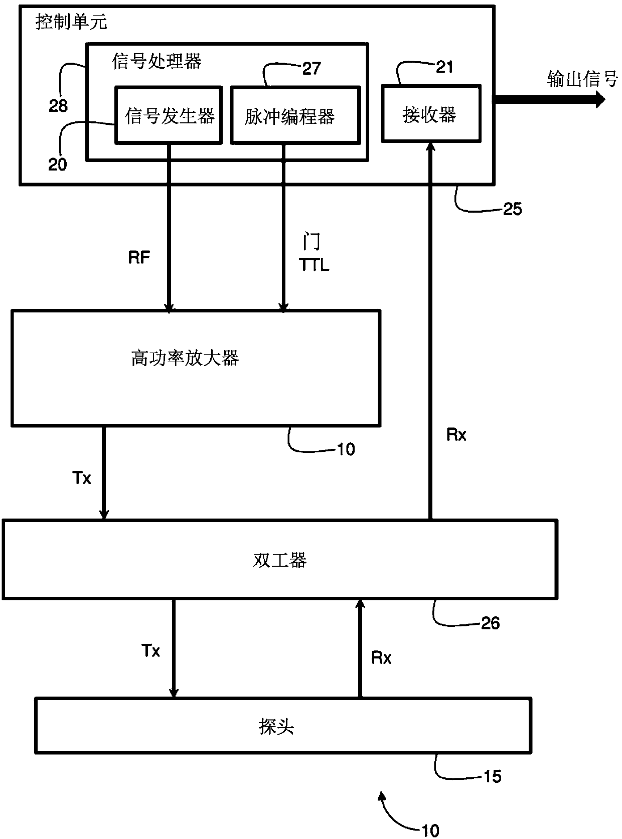
[0018] figure 1 It is a block diagram showing the functions of the system 10 according to the present invention. The system 10 is used to perform non-invasive analysis on the tissue of the subject to distinguish different types of tissues. System 10 includes image 3 with 4 The hand-held probe 15 shown in the picture in the figure has a housing 16 which is formed of a non-ferromagnetic material and has a predetermined cross section at the working end 17 of the housing. The magnetic field source unit 18 in the housing is configured to generate a substantially uniform time-invariant magnetic field within a volume of tissue, the magnetic field having a cross-section equal to the cross-section of the housing at the working end 17 and having a range of 0.05 Tesla to Magnetic field strength in the range of 0.5 Tesla. Provided in the magnetic field source unit 18 is at least one induction coil 19 configured to receive an RF excitation signal (excitation frequency is related to the ma...


PUM

PropertyMeasurementUnit
diameteraaaaaaaaaa
sizeaaaaaaaaaa

Abstract

A system for non-invasive analysis of tissue includes a non-ferromagnetic handheld probe (15) distinguishing between nerves, muscles and blood vessels. A magnetic field source in the probe generates auniform, time-invariant magnetic field within a volume of tissue and contains an inductive coil (19) that receives RF excitation signals in a specific frequency range related to the field strength thereby generating excitation signals perpendicular to a direction of the magnetic field and magnetizing a slice of the living tissue having depth and thickness predetermined by the field strength whereby only living tissue in each slice responds to nuclear spin echo signals. A control unit generates time patterns of the excitation signals and extracts data indicative of the nuclear spin echo signals from the living tissue, determines relaxation curves having time constants T1, T2 and performs statistical processing of the relaxation curves to determine the type of tissue excited by the probe.

Description

Technical field [0001] The invention relates to the use of nuclear magnetic resonance (NMR) to analyze and detect body tissues. Background technique [0002] Nuclear magnetic resonance is a well-known non-invasive method used in medical imaging (MRI). Its effectiveness for radiological diagnosis is the result of the ability to distinguish different types of tissue based on the water content of the tissue in response to different RF pulse sequences. Specifically, the contrast in the MR image is the result of the different relaxation times of the tissue after RF excitation called T1 and T2. [0003] US 2015 / 0018638 provides a technique for non-invasive measurement of blood-related parameters based on an NMR (nuclear) relaxation technique performed using a relatively low constant magnetic field in the range of 0.15 Tesla to 0.5 Tesla. Multiple electromagnetic excitation pulse sequences with relatively low radio frequencies are applied to the living tissue placed in a magnetic field,...

Claims


Application Information

Patent Timeline
Patent Type & Authority Applications(China)
IPC IPC(8): G01R33/44G01R33/38A61B5/055G01N24/08
CPCA61B5/055G01N24/08G01R33/3808G01R33/448A61B5/004
Inventor Y·利维
Owner SHMRI HAIM
Why Eureka
Try Eureka