Automatic splicing method and device for X-ray images, and terminal equipment

An automatic stitching and X-ray technology, applied in the field of image processing, can solve the problems of affecting the quality of image stitching, uneven brightness, difficult to apply to medical X-ray image stitching, etc.

Active Publication Date: 2018-02-06
贵州精准健康数据有限公司
View PDF7 Cites 17 Cited by
  • Summary
  • Abstract
  • Description
  • Claims
  • Application Information

AI Technical Summary

Problems solved by technology

In medical X-ray images, due to the symmetry of the human anatomical structure and the local approximation of the human body structure itself, it is difficult to achieve accurate registration by general methods, which seriously affects the image stitching quality
In addition, compared with other images, medical X-ray images have lower contrast and uneven brightness
Existing digital image stitching methods are difficult to apply to medical X-ray image stitching

Method used


Image

  • Automatic splicing method and device for X-ray images, and terminal equipment
  • Automatic splicing method and device for X-ray images, and terminal equipment
  • Automatic splicing method and device for X-ray images, and terminal equipment

Examples


no. 2 example

[0137] On the basis of the first embodiment of the present invention, before S11, it also includes:

[0138] S10, perform binarization processing on the X-ray images to be stitched.

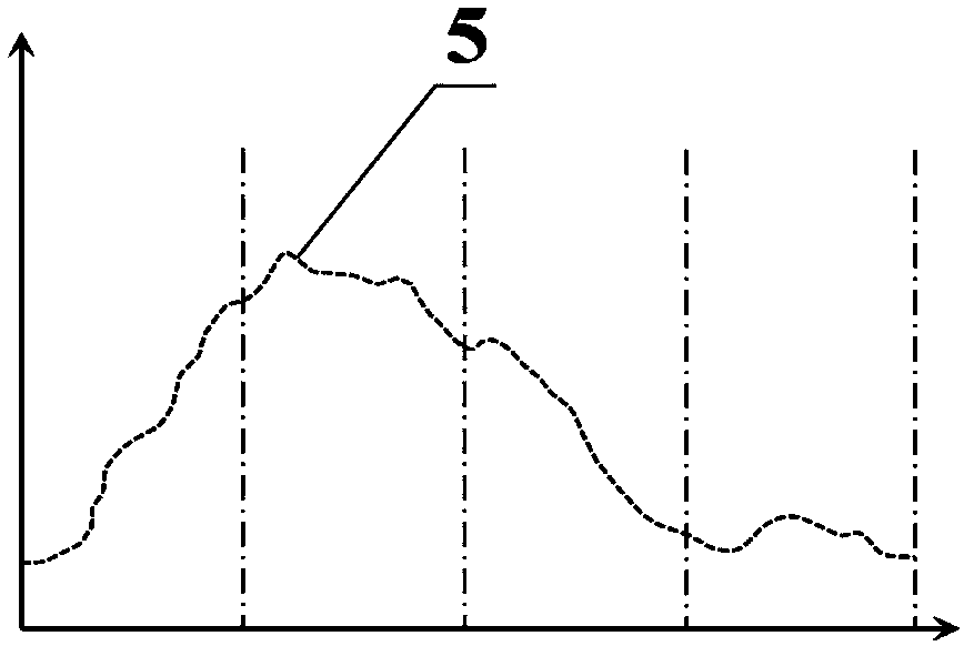
[0139] In the embodiment of the present invention, please refer to Figure 11 , Figure 12 and Figure 13 A schematic diagram of performing binarization processing on the X-ray images to be stitched. The terminal device calculates the grayscale histogram Hist[0] of the X-ray image to be spliced; wherein, the grayscale histogram Hist[0] is a one-dimensional array, and the length of the grayscale histogram Hist[0] L Hist[0] and the maximum gray value G of the X-ray image to be stitched max [0] and the minimum gray value G min [0] has the following relationship: L Hist[0] =G max [0]-G min [0]+1; then count the number of maximum values ​​in the gray histogram Hist[0], and sort according to the size of the maximum values ​​to obtain the gray order set P Hist[0] [0], for computer radiography ...


PUM


Abstract

The invention discloses an automatic splicing method for medical X-ray images. A to-be-spliced X-ray image is subjected to connectivity-based region segmentation, and then a regional image after the region segmentation is obtained. The contour feature points and the skeleton feature points of the regional image are obtained and overlapped to form a seed feature point set. All seed feature points in the seed feature point set are subjected to feature scanning, and then a feature curve is formed. According to the feature curve, the image registration is conducted. The overlapped regions of the regional image after being subjected to image registration are subjected to image fusion and then the image splicing is completed. The invention further provides an automatic splicing device, terminalequipment and a storage medium for medical X-ray images, wherein the characteristics of high precision, fast speed, strong robustness and wide adaptability are realized.

Description

technical field [0001] The invention relates to the field of image processing, in particular to an automatic splicing method, device, terminal equipment and storage medium applied to medical X-ray images. Background technique [0002] X-ray projection imaging technology has the advantages of simple operation and wide projection area at one time, and is one of the most commonly used imaging diagnostic technologies in the medical field. In some X-ray diagnostic scenarios, due to the limitations of the equipment's own hardware, the range of one projection is limited, and it is impossible to capture all medical objects at one time with high resolution and no deformation. For example, in the correction of scoliosis, doctors need to obtain a complete spinal projection image. In order to meet the clinical needs, the part to be photographed needs to be photographed several times, and then the wide-field images are stitched together so that doctors can perform clinical measurements....

Claims


Application Information

Patent Timeline
Patent Type & Authority Applications(China)
IPC IPC(8): G06T5/50G06T7/11G06T7/187G06T7/30
CPCG06T5/50G06T7/11G06T7/187G06T7/30G06T2207/10116
Inventor 孙安玉翟耀斌肖璐吴景姚昌杰陈俊颖许秋娜周丽花
Owner 贵州精准健康数据有限公司
Why Eureka
Try Eureka