Check patentability & draft patents in minutes with Patsnap Eureka AI!

Spatial Mapping Of Molecular Profiles Of Biological Tissue Samples

A tissue sample, two-dimensional space map technology, applied in the determination/inspection of microorganisms, biochemical equipment and methods, etc., can solve problems such as information loss

Active Publication Date: 2018-02-16
KONINKLJIJKE PHILIPS NV
View PDF2 Cites 0 Cited by
  • Summary
  • Abstract
  • Description
  • Claims
  • Application Information

AI Technical Summary

Problems solved by technology

Therefore, by removing the ROI from the slide for further molecular analysis, this information is lost

Method used


Image

  • Spatial Mapping Of Molecular Profiles Of Biological Tissue Samples
  • Spatial Mapping Of Molecular Profiles Of Biological Tissue Samples
  • Spatial Mapping Of Molecular Profiles Of Biological Tissue Samples

Examples


Embodiment

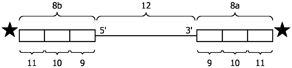
[0184] Types of Protocols for Binding Oligonucleotide Probes to Nucleic Acid Molecules

[0185] .

[0186] Method for Spatial Detection of RNA in FFPE Tissue Samples

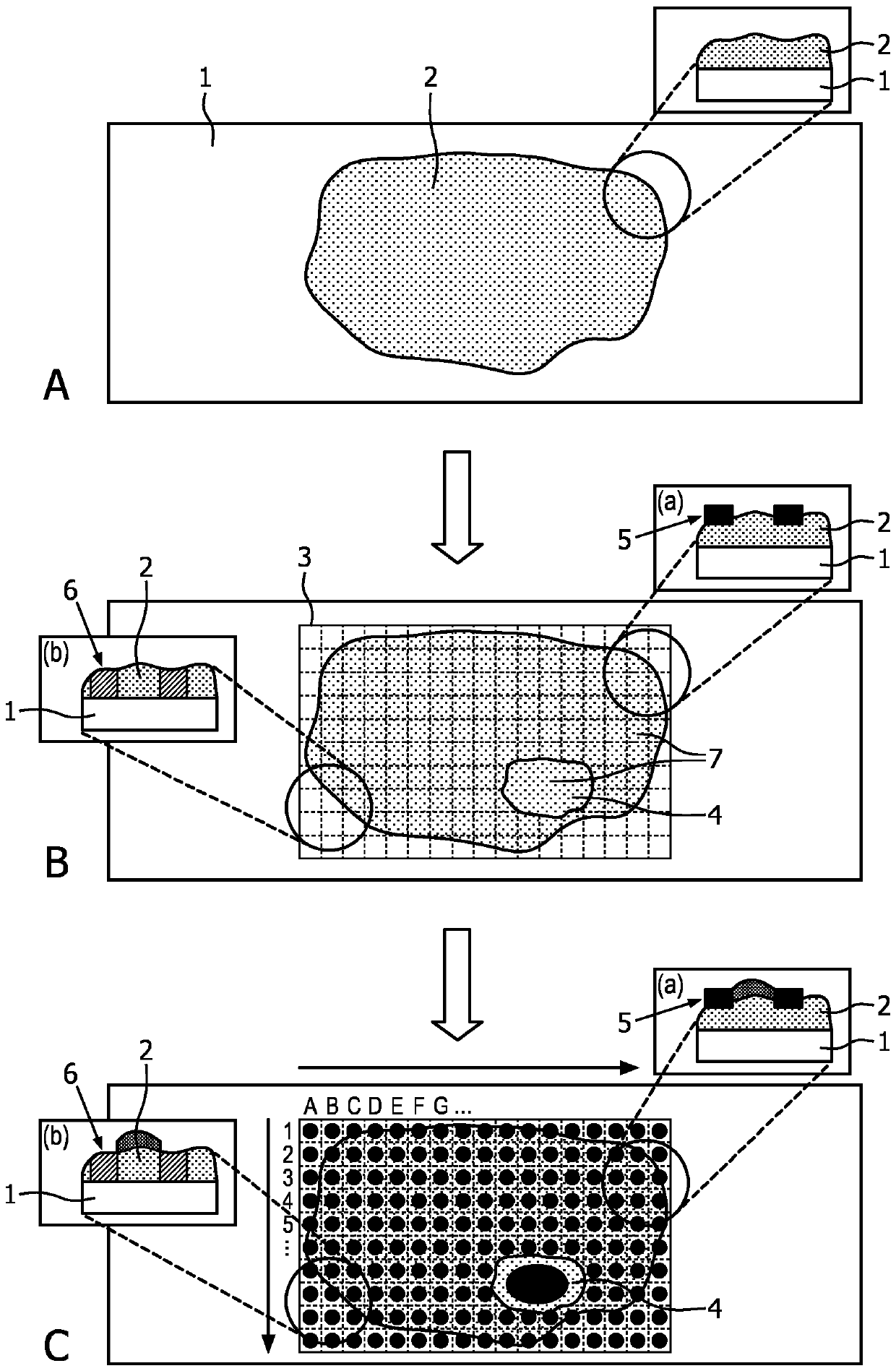
[0187] (Example according to scheme type [C])

[0188] Material: FFPE tissue samples on slides

[0189] Proteinase K (ProtK) was used at 37°C, e.g., as described by Tullis and Rubin (Analytical Biochemistry, 1980, 107(1): 260-264) or according to the manufacturer's instructions using a protein from symbiotic Pleurodenta albicans ( Engyodontium album ) proteinase K (Sigma-Aldrich), followed by heat inactivation of ProtK at 80°C for 15 minutes to treat tissue samples.

[0190] Tissue samples are deparaffinized, eg, using a deparaffinization solution (eg, Qiagen) according to the manufacturer's instructions.

[0191] Images of the entire slide were acquired (Philips Digital Pathology Scanner UFS) and ROIs were identified within the image.

[0192] Different locations were selected within the ROI and a t...


PUM


Abstract

A method is presented that enables the spatial mapping of nucleic acids of tissue samples with high resolution and without sacrificing the degree of multiplexing that is available from next-generationsequencing. The method is based on the application of patterns of barcoded oligonucleotides probes onto predefined locations in a region of interest in a tissue sample. Every nucleic acid analyzed can be allocated to a certain position inside the sample based on the barcode. Various printing technologies can be used and different ways of patterning can be employed, like a regular array with a certain pitch or alternatively an object-based patterning with defined regions of interest without shape constraints.

Description

field of invention [0001] A method that enables spatial mapping of nucleic acids from tissue samples at high resolution is presented. The method is based on the application of patterns of oligonucleotide probes comprising barcode sequences that bind nucleic acids in a sample, taking tissue information into account. Background of the invention [0002] Pathologists are currently performing microscopic examinations of various forms of tissue and cells. Microscopic images reveal information about morphology (eg, cell type, differentiation, etc.) that is the basis for diagnosis. In cases where pathologists want to examine tissues / cells in more detail, biomarker-specific staining protocols are used, such as ER, PR and HER2 in the case of breast cancer. Certain molecular tests are performed on DNA and RNA by so-called in situ hybridization. However, with this method, the number of targets is limited. To improve stratification, perform molecular diagnostic tests to measure gene...

Claims


Application Information

Patent Timeline
IPC IPC(8): C12Q1/6869
CPCC12Q1/6869C12Q2565/519C12Q2543/101C12Q2563/179C12Q2535/122C12Q2535/00C12Q1/6841C12Q1/68C12Q1/6837C12Q1/6844
Inventor M.范德里伊R.维姆伯格-弗里伊德A.皮尔里克
Owner KONINKLJIJKE PHILIPS NV
Features
  • R&D
  • Intellectual Property
  • Life Sciences
  • Materials
  • Tech Scout
  • Unparalleled Data Quality
  • Higher Quality Content
  • 60% Fewer Hallucinations
Learn More