Method, device and computer storage medium for detecting lesion area of ​​breast image

A lesion area and detection method technology, applied in the field of medical image processing, can solve the problems of poor detection result accuracy, irregular shape, and inability to obtain the edge of the mass, and achieve the effect of filtering out false positive lesion areas and improving accuracy

Active Publication Date: 2022-01-28
深圳蓝影医学科技股份有限公司
View PDF0 Cites 0 Cited by
  • Summary
  • Abstract
  • Description
  • Claims
  • Application Information

AI Technical Summary

Problems solved by technology

Among them, the first) method is only based on the feature of breast lesion area density, which is not good for the detection of lesion areas in dense breast images, and the second) method only relies on the K-means clustering algorithm to extract interest Regions, the segmentation effect is better for circular or quasi-circular lesions with clearer edges and more regular shapes, but for lesions with irregular shapes and hidden in dense tissue regions, more accurate tumor edges cannot be obtained.
Therefore, the existing technology cannot accurately segment the lesion area in the dense breast image, resulting in poor detection results

Method used


Image

  • Method, device and computer storage medium for detecting lesion area of ​​breast image
  • Method, device and computer storage medium for detecting lesion area of ​​breast image
  • Method, device and computer storage medium for detecting lesion area of ​​breast image

Examples


Embodiment Construction

[0050] It should be understood that the specific embodiments described here are only used to explain the present invention, not to limit the present invention.

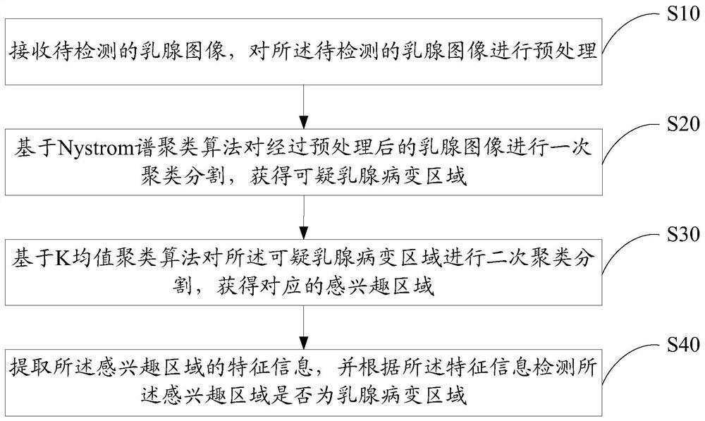
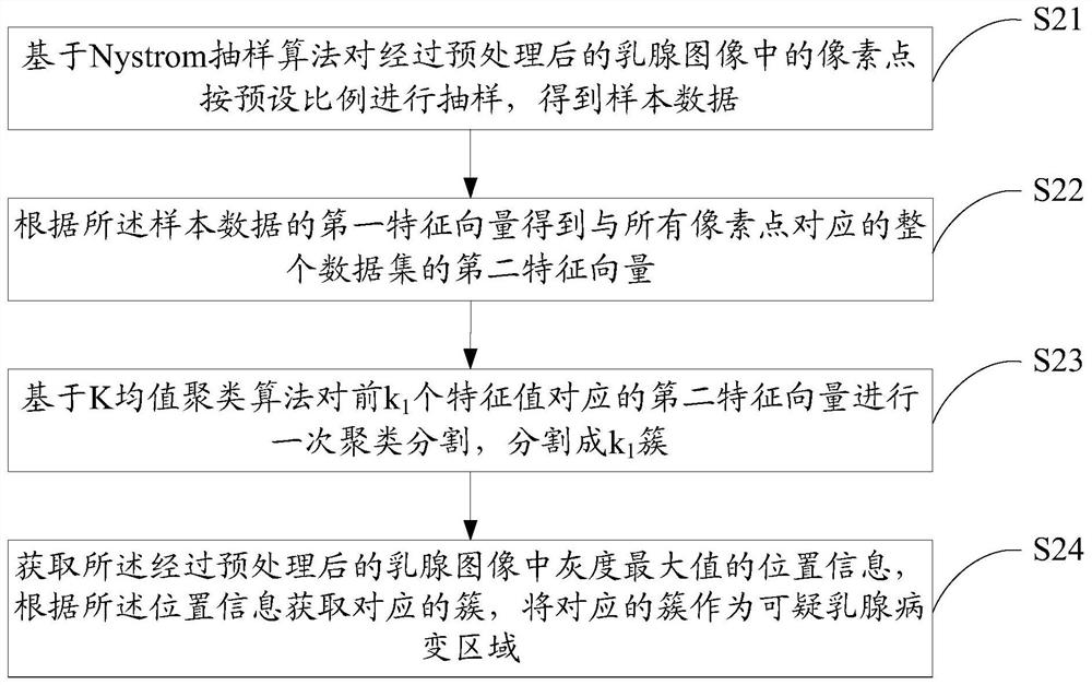
[0051] In the prior art, the processing methods for dense breast images mainly include the following two types: 1) by dividing the dense breast images into several sub-regions, and extracting the density features of each sub-region, performing cluster analysis, and finally displaying Clustering results; 2) Find the region of interest in the mammary image by the K-means method, and then extract features that characterize the mass to distinguish the mass from normal tissue. Among them, the first) method is only based on the feature of breast lesion area density, which is not good for the detection of lesion areas in dense breast images, and the second) method only relies on the K-means clustering algorithm to extract interest Regions, the segmentation effect is better for circular or quasi-circular lesions with clearer ...


PUM


Abstract

The invention discloses a method for detecting lesion areas of mammary gland images. The method includes: receiving a mammary gland image to be detected, and preprocessing the mammary gland image to be detected; performing a clustering and segmentation on the preprocessed mammary gland image based on a Nystrom spectral clustering algorithm to obtain a suspicious mammary gland lesion area; The K-means clustering algorithm performs secondary clustering and segmentation on the suspicious mammary gland lesion region to obtain a corresponding region of interest; extracts feature information of the region of interest, and detects whether the region of interest is Breast lesion area. The invention also discloses a detection device and a computer-readable storage medium for the mammary gland image lesion area. The invention can improve the accuracy of segmenting lesion areas in mammary gland images, thereby improving the accuracy of detection results of mammary gland lesion areas.

Description

technical field [0001] The present invention relates to the technical field of medical image processing, in particular to a method, device and computer storage medium for detecting lesion areas of mammary gland images. Background technique [0002] Breast cancer is a common malignant tumor, early diagnosis and treatment is the key to reduce breast cancer mortality. The computer-aided detection system can help doctors make final diagnostic decisions by detecting suspicious lesion areas, thereby improving the survival rate and quality of life of breast cancer patients. Since lumps and calcification clusters are the most common imaging signs of breast cancer, the automatic detection of lumps and calcifications has also become the two main aspects of the computer-aided diagnosis system. Among them, tumors have always been a difficulty in computer-aided detection due to factors such as blurred edges, various shapes, and low contrast with surrounding tissues. Especially for dens...

Claims


Application Information

Patent Timeline
Patent Type & Authority Patents(China)
IPC IPC(8): G06T7/00G06T7/11G06K9/62
CPCG06T7/0012G06T7/11G06T2207/30068G06F18/2321
Inventor 郑杰胡阳陈晶郭朋
Owner 深圳蓝影医学科技股份有限公司
Why Eureka
Try Eureka