Echocardiographic Ventricle Segmentation Method and Device Based on Deep Learning and Deformable Model

A technology of echocardiography and deformation model, which is applied in the field of medical image processing, and can solve the problems of large manpower, material resources, and negative effects on the calculation of related indicators of the ventricles.

Active Publication Date: 2021-05-28
HARBIN INST OF TECH
View PDF5 Cites 0 Cited by
  • Summary
  • Abstract
  • Description
  • Claims
  • Application Information

AI Technical Summary

Problems solved by technology

[0003] The purpose of the present invention is to solve the disadvantages that the existing manual calibration boundary method usually consumes a lot of manpower and material resources, and the calibration results of different people have certain differences, resulting in a great negative impact on the calculation of ventricle-related indicators. An echocardiographic ventricle cutting method based on deep learning and deformation model is proposed, including:

Method used


Image

  • Echocardiographic Ventricle Segmentation Method and Device Based on Deep Learning and Deformable Model
  • Echocardiographic Ventricle Segmentation Method and Device Based on Deep Learning and Deformable Model
  • Echocardiographic Ventricle Segmentation Method and Device Based on Deep Learning and Deformable Model

Examples


specific Embodiment approach 1

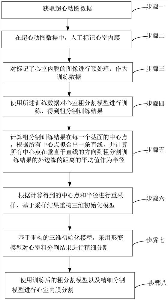
[0034] Specific Embodiment 1: The echocardiographic ventricle segmentation method based on deep learning and deformation model of this embodiment, such as figure 1 shown, including the following steps:

[0035] Step 1: Obtain hypercardiographic data.

[0036] Step 2. Manually mark the endocardium of the ventricles in the hypercardiogram data.

[0037] Step 3: Preprocessing the images marked with the endocardium of the ventricle as training data.

[0038] Step 4: Using the training data to train the rough segmentation model of the ventricle, and obtain the rough segmentation training result.

[0039] Step 5. Calculate the center point of the rough segmentation training result in each section, fit a straight line according to all the center points, and calculate the distance between all the center points and the outer edge of the rough segmentation training result in the direction perpendicular to the straight line Average value as radius.

[0040] Step 6: Perform resampling...

Embodiment approach

[0044] 1. Clinical acquisition of echocardiographic data

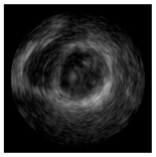
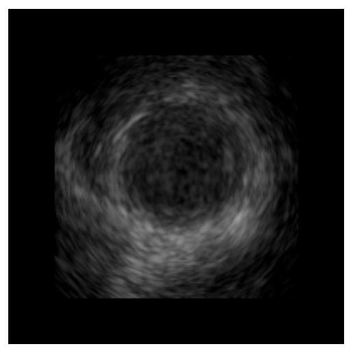
[0045] figure 2 is the echocardiographic short-axis tangential basal image of the ventricle, image 3 is the echocardiographic short-axis tangential mid-portion image of the ventricle, Figure 4 is the echocardiographic ventricular short-axis tangential apical image, Figure 5 is the echocardiographic long-axis "four-chamber heart" slice image, Figure 6 is the echocardiographic long-axis "two-chamber heart" slice image, Figure 7 It's a three-dimensional echocardiogram. according to figure 2 , 3 As shown in , 4, 5, 6 or 7, two-dimensional data or three-dimensional echocardiographic data of each measured person is clinically collected. In order to train a more accurate coarse segmentation model, the number of samples of the tested personnel should be kept above 500 as much as possible. Different tangential data are used as input to provide the heart information of the person being tested, so as to achieve a m...

specific Embodiment approach 2

[0066] Embodiment 2: This embodiment differs from Embodiment 1 in that: the echocardiographic data is two-dimensional, multi-phase and multi-directional echocardiographic slice data or three-dimensional echocardiographic data acquired through ultrasonic equipment.

[0067] Other steps and parameters are the same as those in Embodiment 1.


PUM


Abstract

The present invention relates to an echocardiographic ventricle segmentation method and device based on deep learning and deformation model, which aims to solve the problem that the existing manual boundary calibration usually consumes a lot of manpower and material resources, and the calibration results of different people have certain differences It is proposed due to the shortcomings that have a great negative impact on the calculation of ventricular-related indicators, including: using manually labeled training data to train the ventricular rough segmentation model to obtain the rough segmentation training results; calculating the rough segmentation training results in each A center point of a section, fit a straight line according to all center points, and calculate the average value of the distances from all center points in the direction perpendicular to the line to the outer edge of the rough segmentation training result as the radius; according to the calculated center Points and radii are resampled, and the 3D initialization model is reconstructed based on the sampling results; the deformation model is used to fine-segment the results of the rough segmentation of the ventricle. The present invention is applicable to the image processing of ventricle.

Description

technical field [0001] The invention relates to the field of medical image processing, in particular to a method and device for segmenting the ventricle of an echocardiogram based on deep learning and a deformation model. Background technique [0002] Medical image segmentation, as an important field in the field of medical image processing, is the basis of computer-aided diagnosis and treatment, and is dedicated to efficiently and accurately segmenting organs of interest in medical images. At present, in the field of ventricular segmentation, it mainly relies on manual calibration of the boundaries of the ventricles, and then calculates related indicators of the ventricles based on the calibration results: volume, mass, end-systolic blood volume, end-diastolic blood volume, and ejection fraction of the left and right ventricles. However, such a method of manual boundary calibration usually consumes a lot of manpower and material resources, and the calibration results of dif...

Claims


Application Information

Patent Timeline
Patent Type & Authority Patents(China)
IPC IPC(8): G06T7/11G06T17/00G06N3/04
CPCG06T7/11G06T17/00G06N3/045
Inventor 王宽全董素宇骆功宁袁永峰张恒贵
Owner HARBIN INST OF TECH
Why Eureka
Try Eureka