
Method and device for segmenting left ventricular myocardium

A technology for left ventricle and myocardium, which is applied in the field of image processing and can solve the problems that the filter cannot smooth the curve, cannot effectively suppress the gradient of the non-adventitium boundary, and segment errors.

Active Publication Date: 2019-04-23
SHANGHAI UNITED IMAGING HEALTHCARE
View PDF2 Cites 0 Cited by
  • Summary
  • Abstract
  • Description
  • Claims
  • Application Information

AI Technical Summary

Problems solved by technology

Based on the method of polar coordinate region growth, the Cartesian coordinate image is converted into a polar coordinate image, and then the binary image is obtained by region growth, and finally the Fourier transform is used to smooth the curve. The biggest problem of this method is that region growth is prone to a large number of Over-segmentation, and filtering does not smooth the curve
The method based on dynamic programming uses the gradient of the left ventricle epicardium border and the distance constraint relationship between the borders to search for the left ventricle epicardium border. However, this method cannot effectively suppress the gradient of the non-adventitial border, and it is easy to cause segmentation errors.

Method used

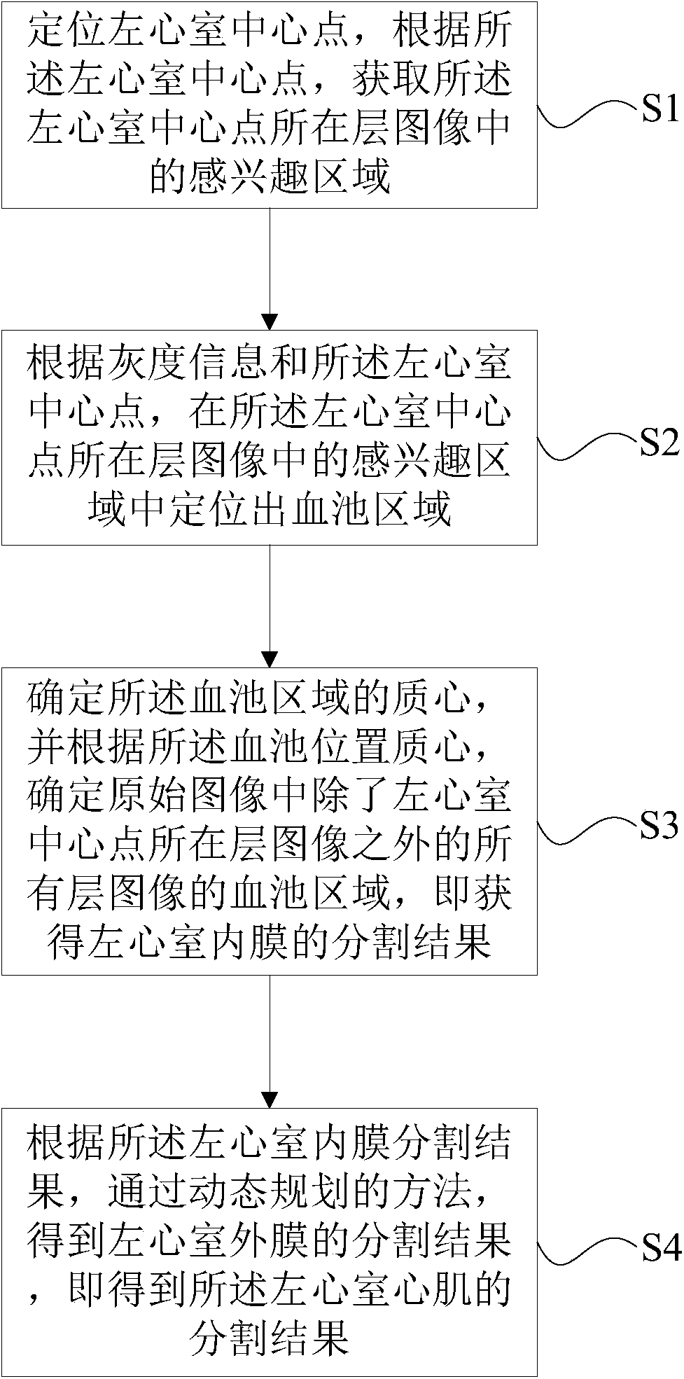
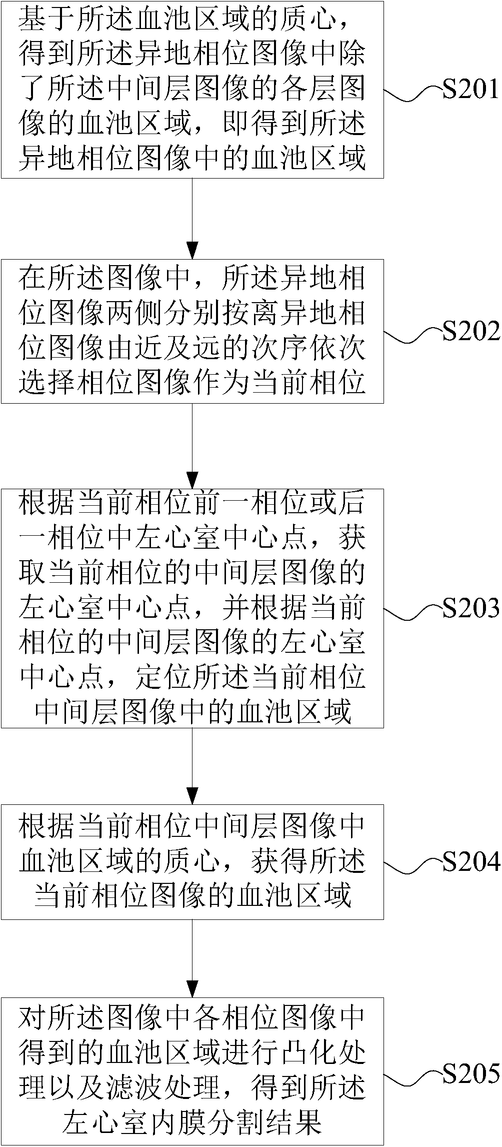
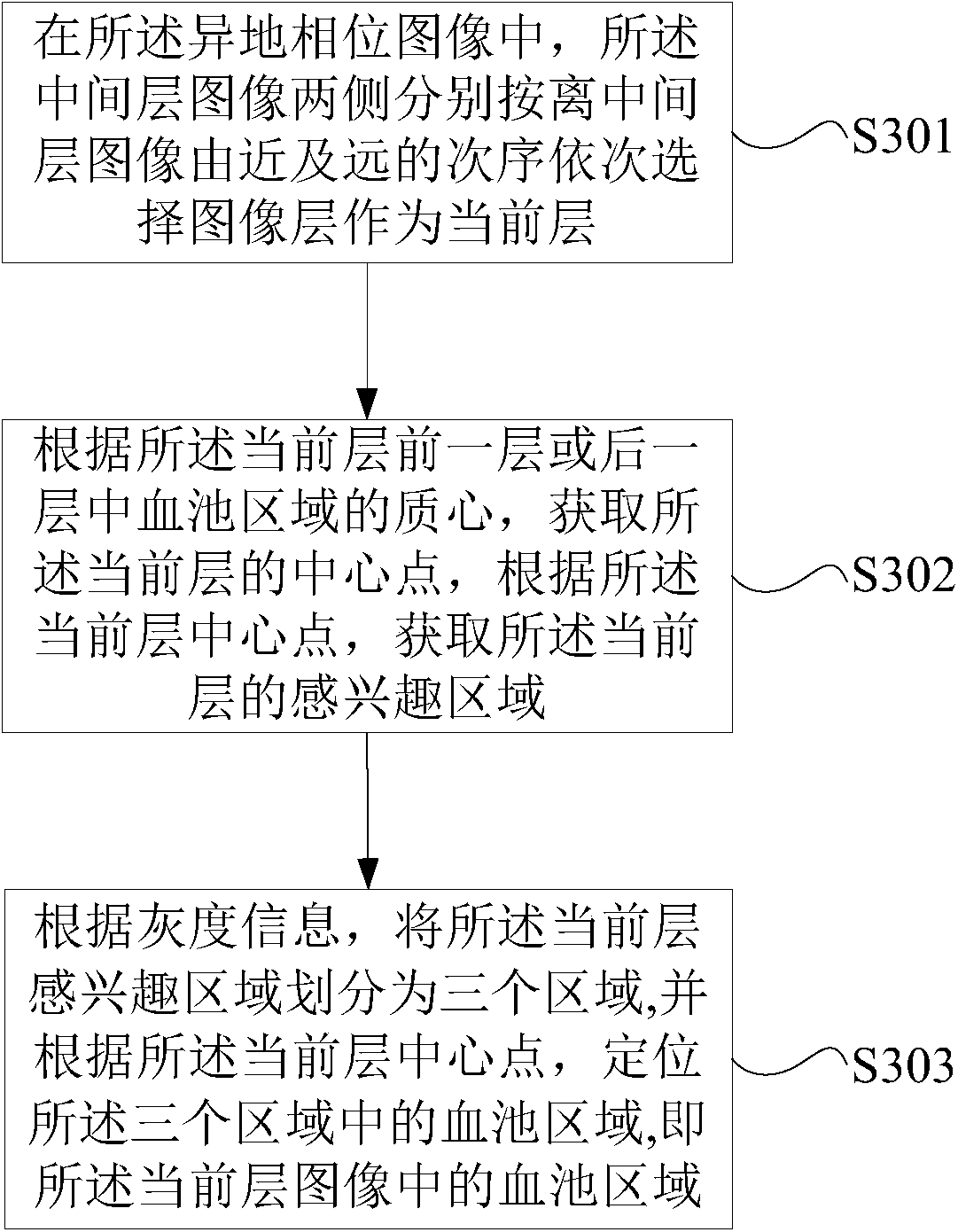

Image

  • Method and device for segmenting left ventricular myocardium
  • Method and device for segmenting left ventricular myocardium
  • Method and device for segmenting left ventricular myocardium

Examples


Embodiment Construction

[0049]In the following description, numerous specific details are set forth in order to provide a thorough understanding of the present invention. However, the present invention can be implemented in many other ways different from those described here, and those skilled in the art can make similar extensions without violating the connotation of the present invention, so the present invention is not limited by the specific implementations disclosed below.

[0050] Secondly, the present invention is described in detail by means of schematic diagrams. When describing the embodiments of the present invention in detail, for convenience of explanation, the schematic diagrams are only examples, which should not limit the protection scope of the present invention.

[0051] A method for segmenting the myocardium of the left ventricle of the present invention will be described in detail below with reference to the drawings and embodiments. The segmentation method of the left ventricular...


PUM


Abstract

The invention provides a division method and device of left ventricular myocardium. The method comprises the following steps of: (1) positioning a left ventricular central point, and acquiring a region of interest in an image at a layer where the left ventricular central point is located; (2) positioning a blood pool region in the region of interest in the image at the layer where the left ventricular central point is located according to grey scale information and the left ventricular central point; (3) determining a mass center of the blood pool region, determining blood pool regions in an original image in all layers except the layer where the left ventricular central point is located, thereby obtaining a division result of left ventricular endocardium; and (4) obtaining a division result of left ventricular adventitia by a dynamic planning method according to the division result of the left ventricular endocardium, and thus obtaining a division result of the left ventricular myocardium. By the technical scheme provided by the invention, the left ventricular myocardium can be fully, uniformly and stably divided.

Description

technical field [0001] The invention relates to the field of image processing, in particular to a method and device for segmenting left ventricular myocardium. Background technique [0002] Segmentation of left ventricular myocardium is the main step in cardiac function analysis. Cardiac function analysis parameters such as left ventricular myocardial wall thickness and ejection volume are all based on accurate segmentation of left ventricular myocardium. The gray scale of the left ventricular myocardium is similar to that of the background, and the papillary muscle and trabeculae in the left ventricle belong to the same substance as the myocardium, and there are artifacts. Therefore, left ventricular myocardium segmentation is a difficult but important task. [0003] At present, there are many methods for segmenting the inner and outer layers of the left ventricle. The segmentation methods of left ventricular endocardium include methods based on active contour shape model...

Claims


Application Information

Patent Timeline
Patent Type & Authority Patents(China)
IPC IPC(8): G06T7/00G06T7/11
Inventor 李强庄吓海钱晓华
Owner SHANGHAI UNITED IMAGING HEALTHCARE
Learn More