
Medical imaging technology evaluation method and device

A technology of medical imaging and evaluation methods, applied in the field of medical imaging

Inactive Publication Date: 2018-06-15
SHANGHAI NEUSOFT MEDICAL TECH LTD
View PDF5 Cites 13 Cited by
  • Summary
  • Abstract
  • Description
  • Claims
  • Application Information

AI Technical Summary

Problems solved by technology

Therefore, existing unilateral methods for evaluating image quality have certain limitations, and this limitation is increasingly emphasized by the academic community

Method used


Image

  • Medical imaging technology evaluation method and device
  • Medical imaging technology evaluation method and device
  • Medical imaging technology evaluation method and device

Examples


Embodiment Construction

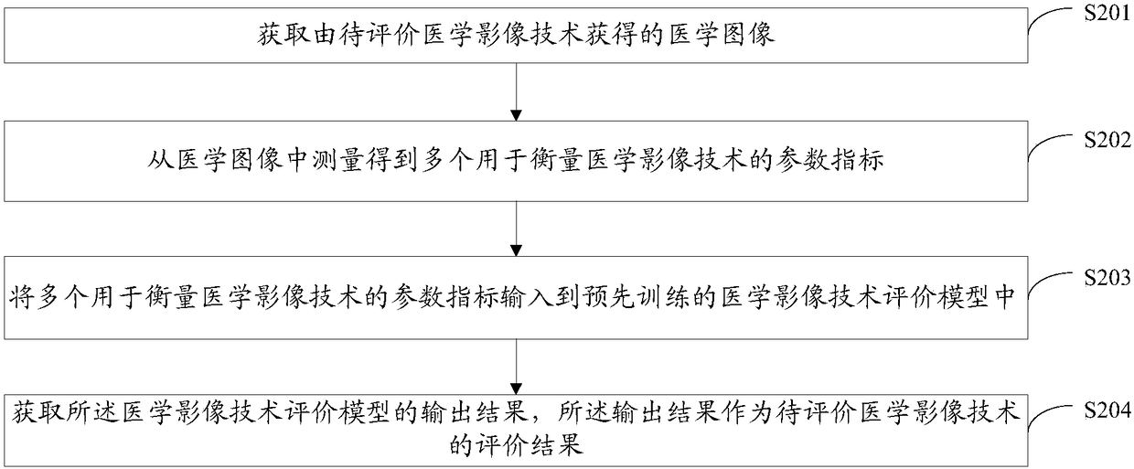
[0049] As described in the background technology, the existing evaluation of medical imaging technology is usually based on the calculation of specific physical quantities, such as the calculation of specific physical quantities such as peak signal-to-noise ratio, mean square error, or entropy. However, due to the imaging principles of medical images and the organization itself The difference in the characteristics of the medical image makes the medical image complex and diverse. On the one hand, this evaluation method based on specific physical quantities is one-sided, and cannot completely reflect the fidelity of the image to the diagnostic information, and cannot give an evaluation with diagnostic value. As a result, it is not accurate as an evaluation standard for measuring medical imaging equipment or methods. On the other hand, this method does not consider the ultimate goal of medical imaging, that is, disease diagnosis, but only from an engineering perspective. It does ...


PUM


Abstract

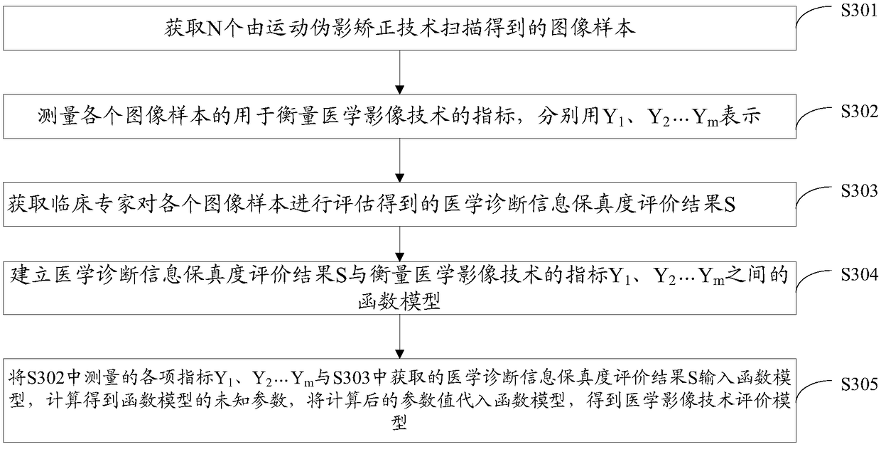
Embodiments of the invention disclose a medical imaging technology evaluation method. The method is used for evaluating a medical imaging technology on the basis of a pre-trained medical imaging technology evaluation model. The medical imaging technology evaluation model is trained according to a plurality of parameter indexes, for measuring the medical imaging technology, of a medical image training sample and a medical diagnosis information fidelity evaluation result of the medical image training sample, so that when the plurality of parameter indexes, for measuring the medical imaging technology, measured from a medical image are input into the medical imaging technology evaluation model, an output result reflecting medical diagnosis information fidelity can be obtained; and the outputresult serves as a medical imaging technology evaluation result and is capable of obtaining an evaluation result reflecting medical diagnosis information fidelity and measuring clinical diagnosis valeof the medical image. The embodiments of the invention furthermore disclose a medical imaging technology evaluation device.

Description

technical field [0001] The present application relates to the field of medical imaging technology, in particular to an evaluation method and device for medical imaging technology. Background technique [0002] Medical imaging is a discipline covering a variety of imaging techniques based on radiodiagnostic medicine. Such as ordinary X-ray photography, digital photography (CR, DR), digital subtraction angiography (DSA), computed tomography (CT), magnetic resonance imaging (MRI), ultrasound technology (B ultrasound, etc.) and radionuclides Scanning (SPECT, PET, etc.) and more. [0003] Due to the differences in signal acquisition and processing methods of imaging equipment and the differences in human visual perception, there are various differences in the finally received medical image information. Therefore, how to comprehensively evaluate medical imaging technologies (including medical Imaging equipment and methods) are crucial to the development of medical imaging. [0...

Claims


Application Information

Patent Timeline
IPC IPC(8): G06K9/62G16H30/20G16H50/20
CPCG06F18/2193G06F18/214
Inventor 陈名亮黄峰
Owner SHANGHAI NEUSOFT MEDICAL TECH LTD
Learn More