

Detachable intravascular imaging catheter and diagnosis system
A technology for imaging catheters and blood vessels, which is applied in the field of intravascular imaging, can solve the problems of high product cost, achieve the effects of simple operation, avoid disinfection process, and facilitate industrialization promotion
- Summary
- Abstract
- Description
- Claims
- Application Information
AI Technical Summary
Problems solved by technology
Method used
Image

Examples
Embodiment 1
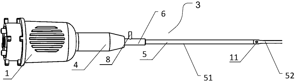
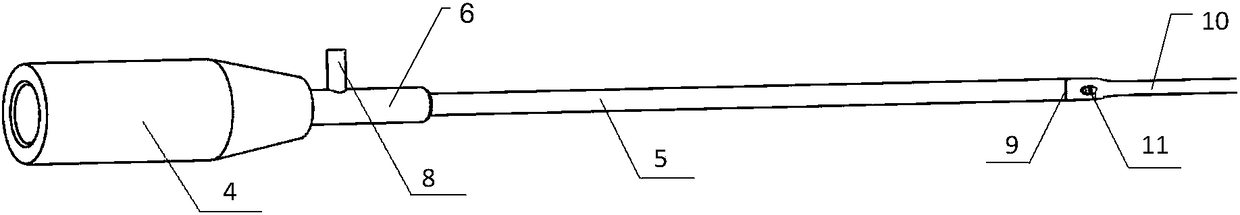
[0034] Such as Figure 1~Figure 5 As shown, a detachable intravascular imaging catheter is connected to the connection port 1. The detachable intravascular imaging catheter includes an imaging inner core 2, an outer sheath 3 and a locking head 4. The outer sheath 3 includes an outer The protective tube 5 and the three-way tube 6, the outer protective tube 5 and the three-way tube 6 are connected, and the outer protective tube 5 includes a proximal area 51 close to the connection port 1 and a part far away from the connection port 1 for guiding into blood vessels. The distal region 52, the proximal region 51 is provided with an imaging lumen, one end of the imaging core 2 is connected to the connection port 1, and the other end of the imaging core 2 is inserted into the imaging tube through the locking head 4 In the lumen; one end of the locking head 4 is connected to the connection port 1, and the other end of the locking head 4 is detachably connected to the outer sheath 3; t...
Embodiment 2
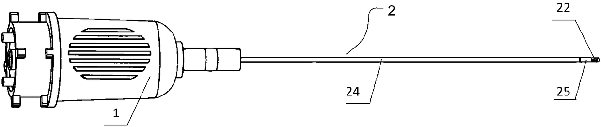
[0039] A diagnostic system such as Figure 6 As shown, it includes a detachable intravascular imaging catheter as described in Embodiment 1, a connection port, a connection unit and a control unit, and the connection port is connected to the imaging unit of the detachable intravascular imaging catheter described in Embodiment 1 and the connection unit, and send the imaging of the target tissue by the imaging unit to the control unit through the connection unit, and the control unit receives, processes and displays the image information.
PUM
 Abstract
Description
Claims
Application Information