
Cerebrovascular segmentation method, system and electronic device

A technology of cerebrovascular and blood vessels, applied in the field of cerebrovascular segmentation methods, systems and electronic equipment, can solve the problems of incomplete vascular network, broken blood vessels, low robustness, etc.

Active Publication Date: 2018-12-28
SHENZHEN INST OF ADVANCED TECH
View PDF8 Cites 10 Cited by
  • Summary
  • Abstract
  • Description
  • Claims
  • Application Information

AI Technical Summary

Problems solved by technology

[0007] However, the existing cerebrovascular segmentation methods still have the following defects: 1), the current method has low robustness, and the extracted blood vessels will have more fractures when the contrast is low; 2), the vascular network extracted by the current method is not complete , thus limiting the scope of clinical diagnosis and treatment

Method used


Image

  • Cerebrovascular segmentation method, system and electronic device
  • Cerebrovascular segmentation method, system and electronic device
  • Cerebrovascular segmentation method, system and electronic device

Examples


Embodiment Construction

[0057] In order to make the purpose, technical solution and advantages of the present application clearer, the present application will be further described in detail below in conjunction with the accompanying drawings and embodiments. It should be understood that the specific embodiments described here are only used to explain the present application, not to limit the present application.

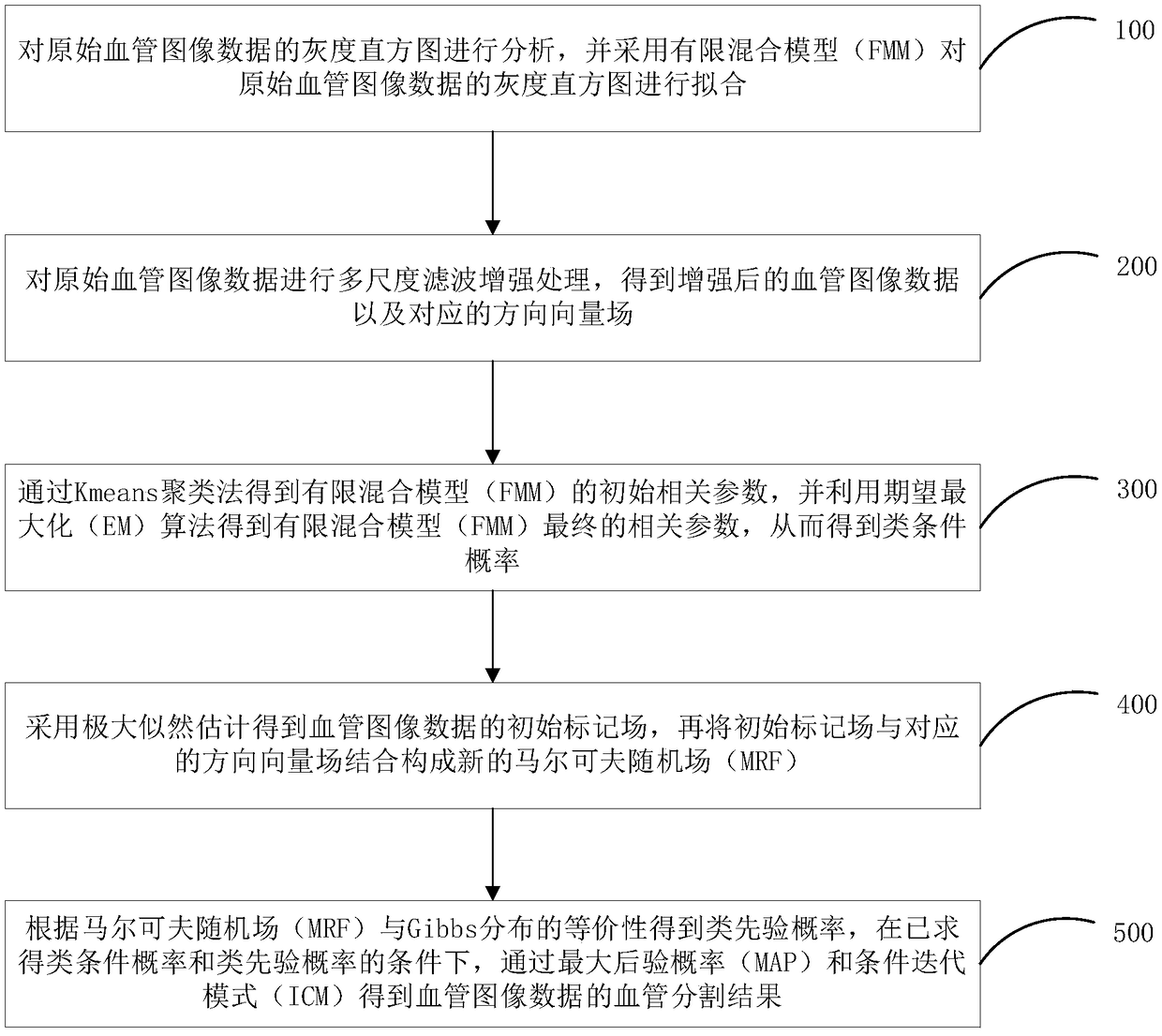
[0058] see figure 1 , is a flow chart of the method for segmenting cerebral blood vessels according to the embodiment of the present application. The cerebrovascular segmentation method of the embodiment of the present application includes the following steps:

[0059] Step 100: analyzing the grayscale histogram of the original blood vessel image data, and fitting the grayscale histogram of the original blood vessel image data by using a finite mixed model (FMM);

[0060] In step 100, the original blood vessel image data is brain TOF-MRA data, specifically, it may also be other types of ...


PUM


Abstract

The present application relates to a cerebral vascular segmentation method, a system and an electronic device. The method comprises the following steps: the original blood vessel image data is processed by multi-scale filtering and enhancement to obtain the enhanced blood vessel image data and the corresponding direction vector field; the finite mixing model is established, and the parameters of the finite mixing model are estimated, and the class conditional probability P (y | x) is obtained; an initial marker field of the blood vessel image data is calculated and the initial marker field iscombined with a corresponding direction vector field to form a new Markov random field; according to the equivalence of Markov random field and Gibbs distribution, a priori-like probability P (x) is obtained; based on the conditional probability P (y | x) and the priori-like probability P (x), the blood vessel segmentation results of the blood vessel image data are obtained through the maximum posterior probability and the conditional iterative mode. The present application can extract more effective blood vessel candidate space, extract blood vessel structure under low contrast, and make theblood vessel network more complete.

Description

technical field [0001] The present application belongs to the technical field of medical image processing, and in particular relates to a method, system and electronic equipment for cerebrovascular segmentation. Background technique [0002] Up to now, a large number of cerebrovascular segmentation methods have been proposed, and some methods have been successfully applied to clinical diagnosis. Existing cerebrovascular segmentation methods are divided into two categories: skeleton-based and non-skeleton. The former segments and reconstructs vessels by first detecting the vessel centerline, while the latter directly extracts 3D vessels. More reports are based on non-skeleton segmentation methods, including: scale analysis method, deformation model method, tracking method, statistical model method and mixed model method. [0003] The blood vessel segmentation problem is regarded as a labeling problem, and all points in the angiographic data are divided into two categories, ...

Claims


Application Information

Patent Timeline
IPC IPC(8): G06T7/11G06T7/143G06T7/194G06T5/00G06K9/62
CPCG06T7/11G06T7/143G06T7/194G06T2207/20024G06T2207/30016G06T2207/30101G06F18/23213G06T5/70
Inventor 李娜周寿军李迟迟王澄
Owner SHENZHEN INST OF ADVANCED TECH
Learn More