Preparation method of scanning electron microscope sample of bivalve shell

A scanning electron microscope, bivalve technology, applied in the field of biology and materials science, to achieve the effect of easy access, quick and simple operation, and easy to use

Active Publication Date: 2019-01-08
NANJING UNIV OF SCI & TECH
View PDF3 Cites 1 Cited by
  • Summary
  • Abstract
  • Description
  • Claims
  • Application Information

AI Technical Summary

Problems solved by technology

[0005] The purpose of the present invention is mainly aimed at the problem of the preparation of the...

Method used


Image

  • Preparation method of scanning electron microscope sample of bivalve shell
  • Preparation method of scanning electron microscope sample of bivalve shell
  • Preparation method of scanning electron microscope sample of bivalve shell

Examples


Embodiment 1

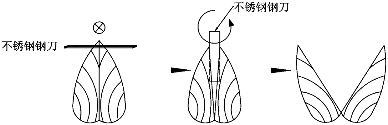
[0039] Embodiment 1: In the following practical example, the fresh non-deactivated triangular sail clam shells of 2-3 years old in Zhuji, Zhejiang are taken as the experimental object, and the structure needs to be photographed with a scanning electron microscope, and the single-layer aragonite wafer in the nacre layer on the 30 growth line The thickness is measured. In conjunction with the accompanying drawings, this example is used to further illustrate the present invention, but the present invention is not limited thereto.

[0040] Firstly, pre-treatment and cleaning are carried out on the triangular sail clam shells:

[0041] 1) if figure 1 As shown, in order to facilitate the insertion of the stainless steel knife, use a stainless steel knife to cut a flat fracture parallel to the closed edge of the triangle sail clam shell, then insert the knife head perpendicular to the flat fracture into the shell, and rotate the stainless steel knife left and right to make the trian...

Embodiment 2

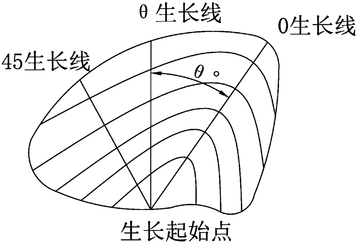
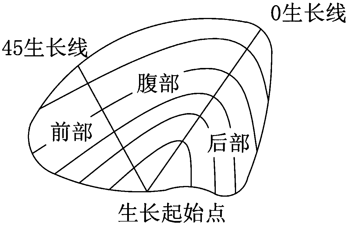
[0055] Example 2: In the following actual example, the air-dried inactivated mother-of-pearl shells aged 2-3 years in Zhuji, Zhejiang Province were used as the experimental object. It was necessary to take a scanning electron microscope to observe the structure and study the thickness difference of the nacre arsonite wafers on the 0 growth line. In conjunction with the accompanying drawings, this example is used to further illustrate the present invention, but the present invention is not limited thereto.

[0056] like figure 2 As shown, put the air-dried shells on the table with the top facing up, fix one end of the soft thin wire to the starting point of the shell, and then rotate the other end along the outer edge of the shell until you find the point farthest from the starting point of the shell. At one point, use a marker pen to draw a curve along the surface of the shell to get the 0 growth line of the shell.

[0057] With the 0 growth line as the center line, draw two...


PUM

PropertyMeasurementUnit
Particle sizeaaaaaaaaaa
Thicknessaaaaaaaaaa
Particle sizeaaaaaaaaaa

Abstract

The invention relates to the fields of biology and material science, in particular to a preparation method of a scanning electron microscope sample of a bivalve shell. The method comprises the following four parts of pre-processing, positioning, sampling and post-processing. The preparation method of the scanning electron microscope sample of the bivalve shell is provided by utilizing common experiment equipments in a laboratory. By adopting the method provided by the invention, the scanning electron microscope sample of the bivalve shell can be prepared quickly, simply and efficiently, a clear scanning electron microscope picture of the bivalve shell can be effectively obtained.

Description

technical field [0001] The invention relates to the fields of biology and material science, in particular to a method for preparing a bivalve shell for a scanning electron microscope sample. Background technique [0002] With the development of social economy and the change of the use environment, the lack of mechanical properties and strength of traditional structural materials limits its application in wider fields. Therefore, constructing structural materials with special structures to enhance their mechanical properties has attracted increasing attention. [0003] Under the natural law of the survival of the fittest, in order to adapt to changes in the external environment, natural biological materials have undergone continuous evolution for hundreds of millions of years or even longer, and their structures and various properties have gradually become perfect. As a typical natural biological material, the bivalve shell is also the most important freshwater pearl mussel ...

Claims


Application Information

Patent Timeline
IPC IPC(8): G01N23/2202
CPCG01N23/2202
Inventor 周琦邵浩彬王克鸿朱军雷济旭徐俊强郭顺
Owner NANJING UNIV OF SCI & TECH
Why Eureka
Try Eureka