Image processor, image processing method, and image processing program
An image processing device and image processing technology, applied in image data processing, image enhancement, image analysis and other directions, can solve problems such as being unsuitable for health diagnosis purposes, judgment, and inability to detect
- Summary
- Abstract
- Description
- Claims
- Application Information
AI Technical Summary
Problems solved by technology
Method used
Image
Examples
Deformed example 1
[0106] Figure 7 It is a figure which shows an example of the recognizer M concerning the modification 1.
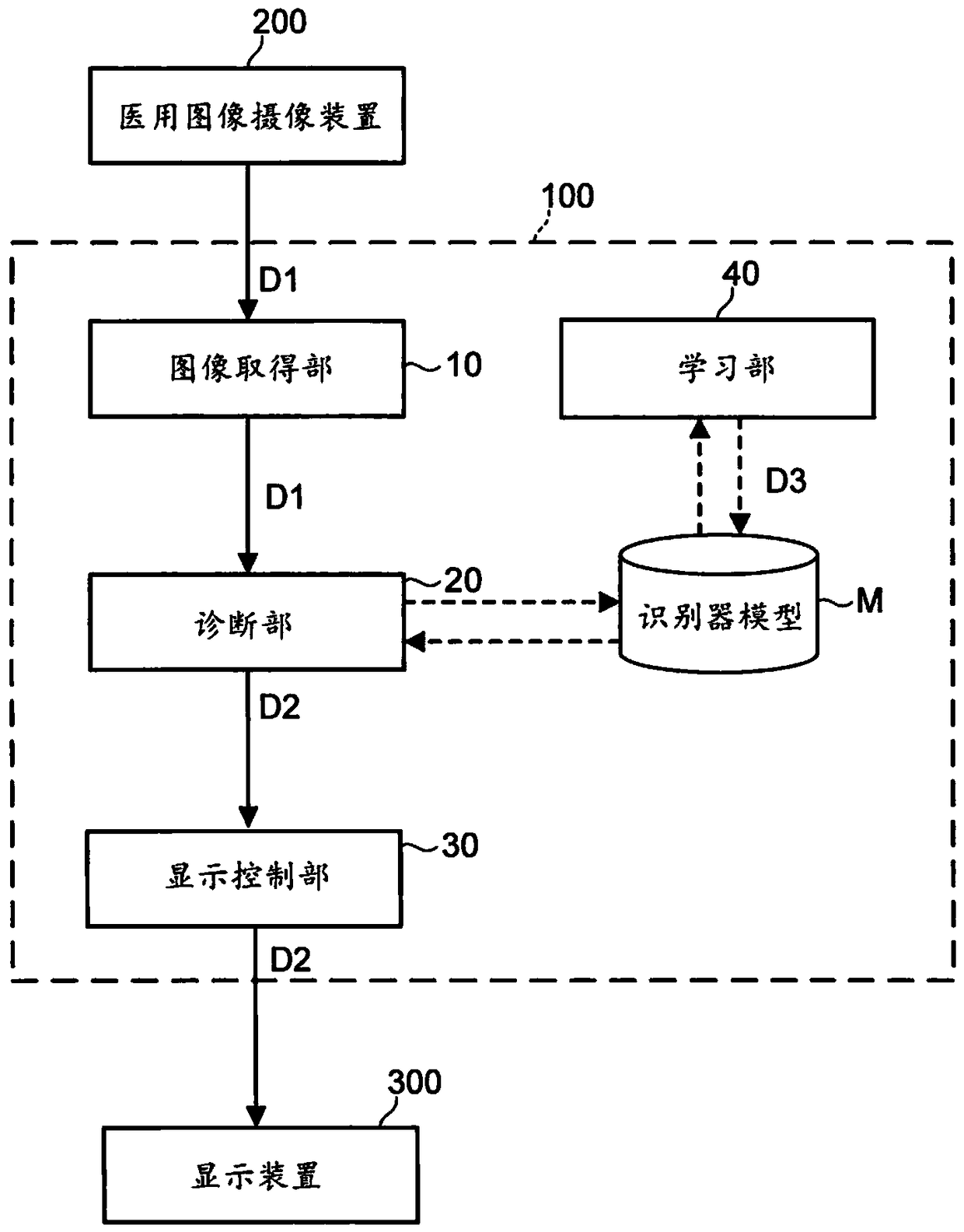
[0107] The diagnostic unit 20 according to Modification 1 differs from the above in that it divides the entire image area of a medical image into a plurality of image areas (here, divided into 9 parts D1a to D1i) and calculates the degree of normality for each image area. The implementation is different.
[0108] The aspect related to Modification 1 can be realized, for example, by providing a classifier M for performing image analysis for each image region of a medical image. exist Figure 7 , there are 9 different classifiers Ma~Mi provided to correspond to the 9 image regions D1a~D1i respectively. In addition, the classifier M for performing image analysis may be provided for each internal organ part of the medical image.
[0109] The display control unit 30 according to Modification 1, for example, associates the degree of normality calculated for each image re...
Deformed example 2
[0113] Figure 8 It is a figure which shows an example of the identifier M concerning the modification 2.
[0114] The diagnosis unit 20 according to this modification 2 is different from the above-mentioned embodiment in that it calculates the normality for each pixel region of a medical image (representing a region forming one pixel or a region of a plurality of pixels forming one division. The same applies hereinafter). different ways.
[0115] The aspect of Modification 2 can be realized, for example, by providing an output element for each pixel area of a medical image in the recognition unit Nb of CNN (also referred to as R-CNN).
[0116] The display control unit 30 according to Modification 2, for example, correlates the normality of each pixel region with the position of the pixel region in the medical image, and displays it on the display device 300 . At this time, the display control unit 30 expresses, for example, the normality conversion of each pixel area as c...
Deformed example 3
[0120] The image processing device 100 according to Modification 3 differs from the above-described embodiment in the configuration of the display control unit 30 .
[0121] The display control unit 30 sets the order in which the plurality of medical images are displayed on the display device 300 based on the respective normality degrees of the plurality of medical images, for example, after calculating the normality of the plurality of medical images. Then, the display control unit 30 outputs, for example, the medical image data D1 and the normality data D2 to the display device 300 in a set order.
[0122] In this way, for example, among a plurality of medical images, those with a higher possibility of being in an abnormal state are displayed on the display device 300 in order, so that a subject with a high necessity or urgency can receive a formal diagnosis by a doctor or the like. .
[0123] In addition, the display control unit 30 may set whether to display a plurality o...
PUM
 Abstract
Description
Claims
Application Information
 - R&D Engineer
- R&D Manager
- IP Professional
- Industry Leading Data Capabilities
- Powerful AI technology
- Patent DNA Extraction
Browse by: Latest US Patents, China's latest patents, Technical Efficacy Thesaurus, Application Domain, Technology Topic.
© 2024 PatSnap. All rights reserved.Legal|Privacy policy|Modern Slavery Act Transparency Statement|Sitemap


