Brain perfusion image segmentation method, device, server and storage medium

An image segmentation and cerebral perfusion technology, applied in the field of medical image analysis, can solve the problems of poor segmentation effect, ignoring cerebrospinal fluid and blood vessel neighborhood information, etc., and achieve the effect of accurate segmentation

Active Publication Date: 2022-01-11
SHANGHAI UNITED IMAGING HEALTHCARE
View PDF11 Cites 0 Cited by
  • Summary
  • Abstract
  • Description
  • Claims
  • Application Information

AI Technical Summary

Problems solved by technology

In the above method, the cerebrospinal fluid and blood vessels are directly segmented through the threshold, but the neighborhood information of cerebrospinal fluid and blood vessels is ignored, and the segmentation effect is not good.

Method used


Image

  • Brain perfusion image segmentation method, device, server and storage medium
  • Brain perfusion image segmentation method, device, server and storage medium
  • Brain perfusion image segmentation method, device, server and storage medium

Examples


Embodiment 1

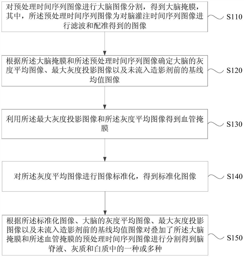
[0087] figure 1 It is a flow chart of the brain perfusion image segmentation method provided by Embodiment 1 of the present invention. This embodiment is applicable to the situation of segmenting the brain perfusion image. This method can be executed by a brain perfusion image segmentation device, which can be configured, for example, in in the server. Such as figure 1 As shown, the method specifically includes:

[0088] S110. Perform brain image segmentation on the pre-processed time-series images to obtain a brain mask, wherein the pre-processed time-series images are images obtained by filtering and registering the brain perfusion time-series images.

[0089] Specifically, first obtain the time-series images of cerebral perfusion, and preprocess the time-series images of cerebral perfusion to obtain the pre-processed time-series images, wherein the time-series images of cerebral perfusion include brain perfusion images collected at various time points . The brain perfus...

Embodiment 2

[0131] figure 2 It is a flow chart of the cerebral perfusion image segmentation method provided by the second embodiment of the present invention. The second embodiment further illustrates the specific method for segmenting the cerebrospinal fluid on the basis of the first embodiment. Such as figure 2 As shown, the brain perfusion image segmentation methods include:

[0132] S210. Perform brain image segmentation on the pre-processed time-series images to obtain a brain mask, wherein the pre-processed time-series images are images obtained by filtering and registering the brain perfusion time-series images.

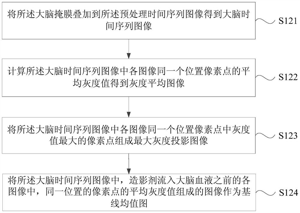
[0133] S220. Determine, according to the brain mask and the preprocessed time-series images, a gray-scale average image, a maximum gray-scale projection image, and a baseline average image before contrast agent flows.

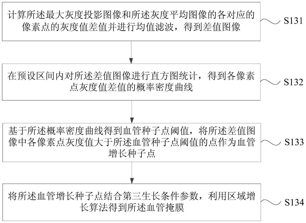
[0134] S230. Obtain a blood vessel mask by using the maximum grayscale projection image and the grayscale average image.

[0135] S240. Perform image...

Embodiment 3

[0145] image 3 It is a flow chart of the brain perfusion image segmentation method provided in Embodiment 3 of the present invention. In Embodiment 3, gray matter and white matter regions are further segmented on the basis of Embodiment 1 and Embodiment 2. Such as image 3 As shown, gray matter segmentation methods include:

[0146] S310. Calculate the distribution probability curve of the gray value of the standardized image, and use a three-Gaussian mixture model to perform fitting to obtain a fitting result.

[0147] S320. Calculate the gray matter seed point threshold according to the fitting result.

[0148] S330. In the preprocessed time-series images superimposed with the brain mask, blood vessel mask and cerebrospinal fluid mask, determine that the pixel points whose gray value is greater than the gray matter seed point threshold are gray matter seed points.

[0149] Specifically, the brain mask, blood vessel mask, and cerebrospinal fluid mask are superimposed on t...


PUM


Abstract

The embodiment of the invention discloses a brain perfusion image segmentation method, device, server and storage medium. The method comprises: performing brain image segmentation on the preprocessed time series images to obtain a brain mask; determining a feature image according to the brain mask and the preprocessed time series images; using the maximum grayscale projection image and the grayscale average image in the feature image to obtain the blood vessel mask The average gray image was normalized to obtain a standardized image; the brain mask and blood vessel mask were superimposed according to the normalized image, the gray average image of the brain, the maximum gray projection image, and the baseline average image before the contrast agent flowed into it. The preprocessed time series images of the membrane are segmented to obtain one or more of cerebrospinal fluid, gray matter and white matter. The embodiment of the present invention solves the problem of poor edge segmentation of different brain tissues in the brain perfusion image segmentation in the prior art, and realizes precise segmentation of different brain tissues in the brain perfusion image and automation of image processing.

Description

technical field [0001] Embodiments of the present invention relate to a medical image analysis technology, in particular to a brain perfusion image segmentation method, device, server and storage medium. Background technique [0002] Cerebral perfusion imaging is a technology that injects radiopharmaceuticals that can pass through the normal blood-brain barrier into the human body, obtains brain perfusion images through imaging equipment, and uses brain perfusion images to diagnose brain diseases. Specifically, in the diagnosis process, it is necessary to segment the brain perfusion image to obtain the distribution of different tissues and determine the lesion location of the patient's brain tissue. [0003] In the prior art, the method of segmenting the brain perfusion image is: segment the skull based on the scheme of the brain perfusion image atlas, then use the threshold method to segment the cerebrospinal fluid and blood vessels, and finally construct the feature vector...

Claims


Application Information

Patent Timeline
Patent Type & Authority Patents(China)
IPC IPC(8): G06T7/11G06T7/136G06T7/194
CPCG06T7/11G06T7/136G06T7/194G06T2207/30016G06T2207/30104
Inventor 龚震寰
Owner SHANGHAI UNITED IMAGING HEALTHCARE
Why Eureka
Try Eureka