Brain perfusion image segmentation method and device, server and storage medium
An image segmentation and cerebral perfusion technology, applied in the field of medical image analysis, can solve the problems of poor segmentation effect, ignoring cerebrospinal fluid and blood vessel neighborhood information, etc.
- Summary
- Abstract
- Description
- Claims
- Application Information
AI Technical Summary
Problems solved by technology
Method used
Image
Examples
Embodiment 1
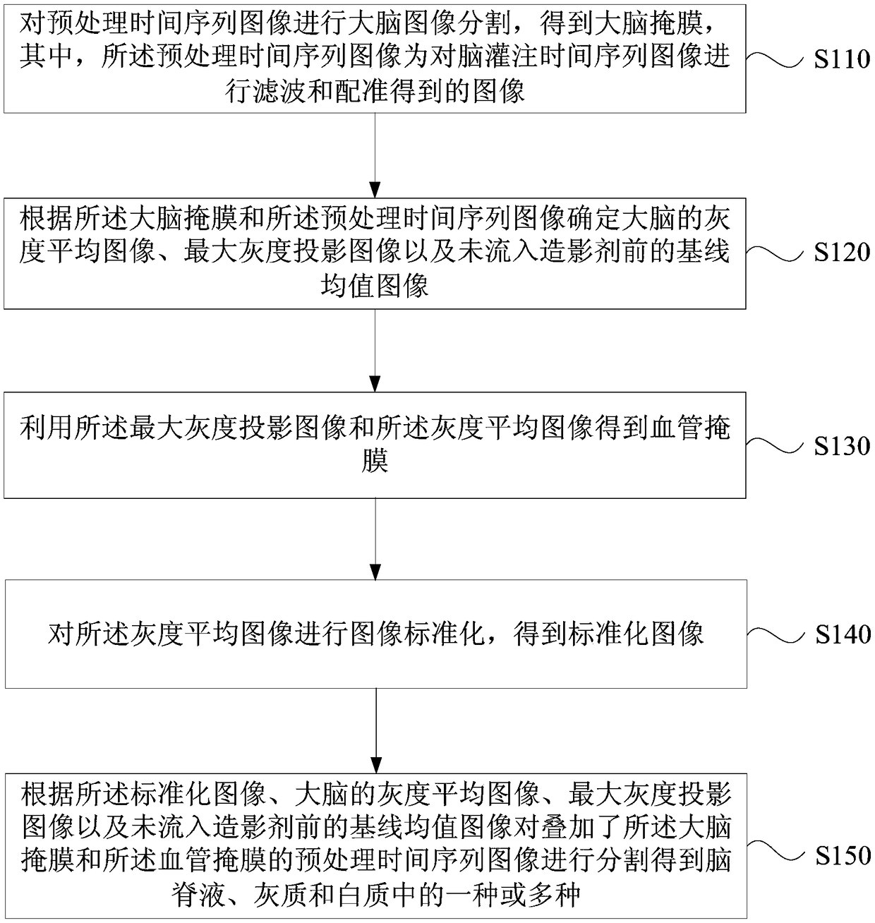
[0087] figure 1 It is a flow chart of the brain perfusion image segmentation method provided by Embodiment 1 of the present invention. This embodiment is applicable to the situation of segmenting the brain perfusion image. This method can be executed by a brain perfusion image segmentation device, which can be configured, for example, in in the server. like figure 1 As shown, the method specifically includes:
[0088] S110. Perform brain image segmentation on the pre-processed time-series images to obtain a brain mask, wherein the pre-processed time-series images are images obtained by filtering and registering the brain perfusion time-series images.
[0089] Specifically, first obtain the time-series images of cerebral perfusion, and preprocess the time-series images of cerebral perfusion to obtain the pre-processed time-series images, wherein the time-series images of cerebral perfusion include brain perfusion images collected at various time points . The brain perfusion...
Embodiment 2
[0131] figure 2 It is a flow chart of the cerebral perfusion image segmentation method provided by the second embodiment of the present invention. The second embodiment further illustrates the specific method for segmenting the cerebrospinal fluid on the basis of the first embodiment. like figure 2 As shown, the brain perfusion image segmentation methods include:
[0132] S210. Perform brain image segmentation on the pre-processed time-series images to obtain a brain mask, wherein the pre-processed time-series images are images obtained by filtering and registering the brain perfusion time-series images.
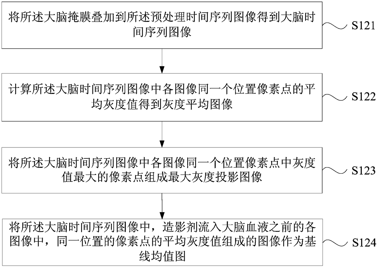
[0133] S220. Determine, according to the brain mask and the preprocessed time-series images, a gray-scale average image, a maximum gray-scale projection image, and a baseline average image before contrast agent flows.
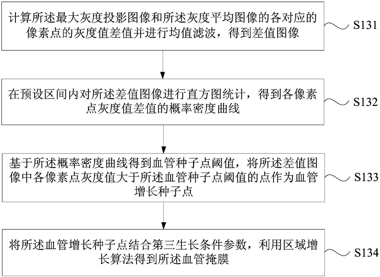
[0134] S230. Obtain a blood vessel mask by using the maximum grayscale projection image and the grayscale average image.
[0135] S240. Perform image no...
Embodiment 3
[0145] image 3 It is a flow chart of the brain perfusion image segmentation method provided in Embodiment 3 of the present invention. In Embodiment 3, gray matter and white matter regions are further segmented on the basis of Embodiment 1 and Embodiment 2. like image 3 As shown, gray matter segmentation methods include:
[0146] S310. Calculate the distribution probability curve of the gray value of the standardized image, and use a three-Gaussian mixture model to perform fitting to obtain a fitting result.
[0147] S320. Calculate the gray matter seed point threshold according to the fitting result.
[0148] S330. In the preprocessed time-series images superimposed with the brain mask, blood vessel mask and cerebrospinal fluid mask, determine that the pixel points whose gray value is greater than the gray matter seed point threshold are gray matter seed points.
[0149] Specifically, the brain mask, blood vessel mask, and cerebrospinal fluid mask are superimposed on the ...
PUM
 Abstract
Description
Claims
Application Information
 - R&D
- Intellectual Property
- Life Sciences
- Materials
- Tech Scout
- Unparalleled Data Quality
- Higher Quality Content
- 60% Fewer Hallucinations
Browse by: Latest US Patents, China's latest patents, Technical Efficacy Thesaurus, Application Domain, Technology Topic, Popular Technical Reports.
© 2025 PatSnap. All rights reserved.Legal|Privacy policy|Modern Slavery Act Transparency Statement|Sitemap|About US| Contact US: help@patsnap.com


