
An automatic medical image segmentation method based on multi-atlas label fusion

A label fusion, medical image technology, applied in image analysis, image enhancement, image data processing and other directions, can solve the problem of reduced accuracy of fusion results, failure to consider local similarity information between target images and atlas grayscale images, and pixel label assignment errors. And other issues

Inactive Publication Date: 2019-03-26
HARBIN UNIV OF SCI & TECH
View PDF5 Cites 8 Cited by
  • Summary
  • Abstract
  • Description
  • Claims
  • Application Information

AI Technical Summary

Problems solved by technology

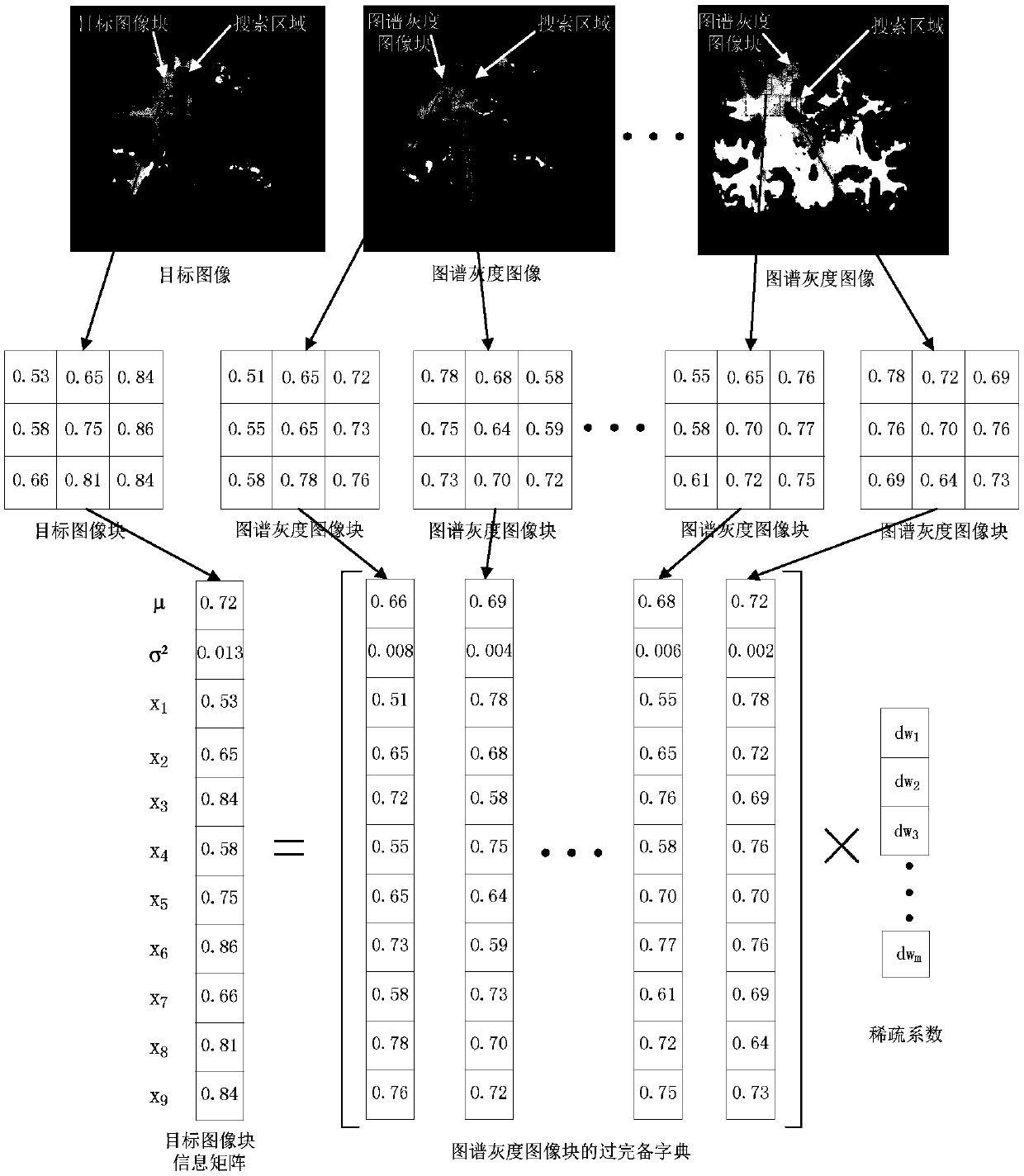
However, the atlas label fusion method based on sparse representation only considers the information of the corresponding pixels between the target image and the atlas gray image, and does not consider the local similarity information between the target image and the atlas gray image, which leads to a low similarity with the icon image. Atlas images will also be fused, resulting in reduced accuracy of fusion results
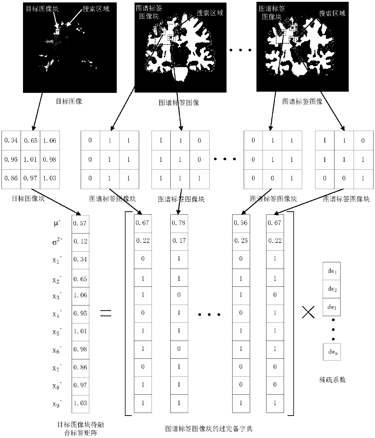
The map label fusion method based on sparse representation uses a fixed threshold to binarize the map label fusion results and assign labels to each pixel, but the mean value of the map label fusion results of different target tissues is different, even for the same target. The results of map label fusion in different regions are also quite different, so using a fixed threshold to binarize the result of map label fusion will cause some pixel label assignment errors

Method used


Image

  • An automatic medical image segmentation method based on multi-atlas label fusion
  • An automatic medical image segmentation method based on multi-atlas label fusion
  • An automatic medical image segmentation method based on multi-atlas label fusion

Examples


Embodiment Construction

[0053] In this application, an MR image of a human brain is used as a segmentation object for description. It should be understood that the present invention is not limited to application to MR images of the human brain.

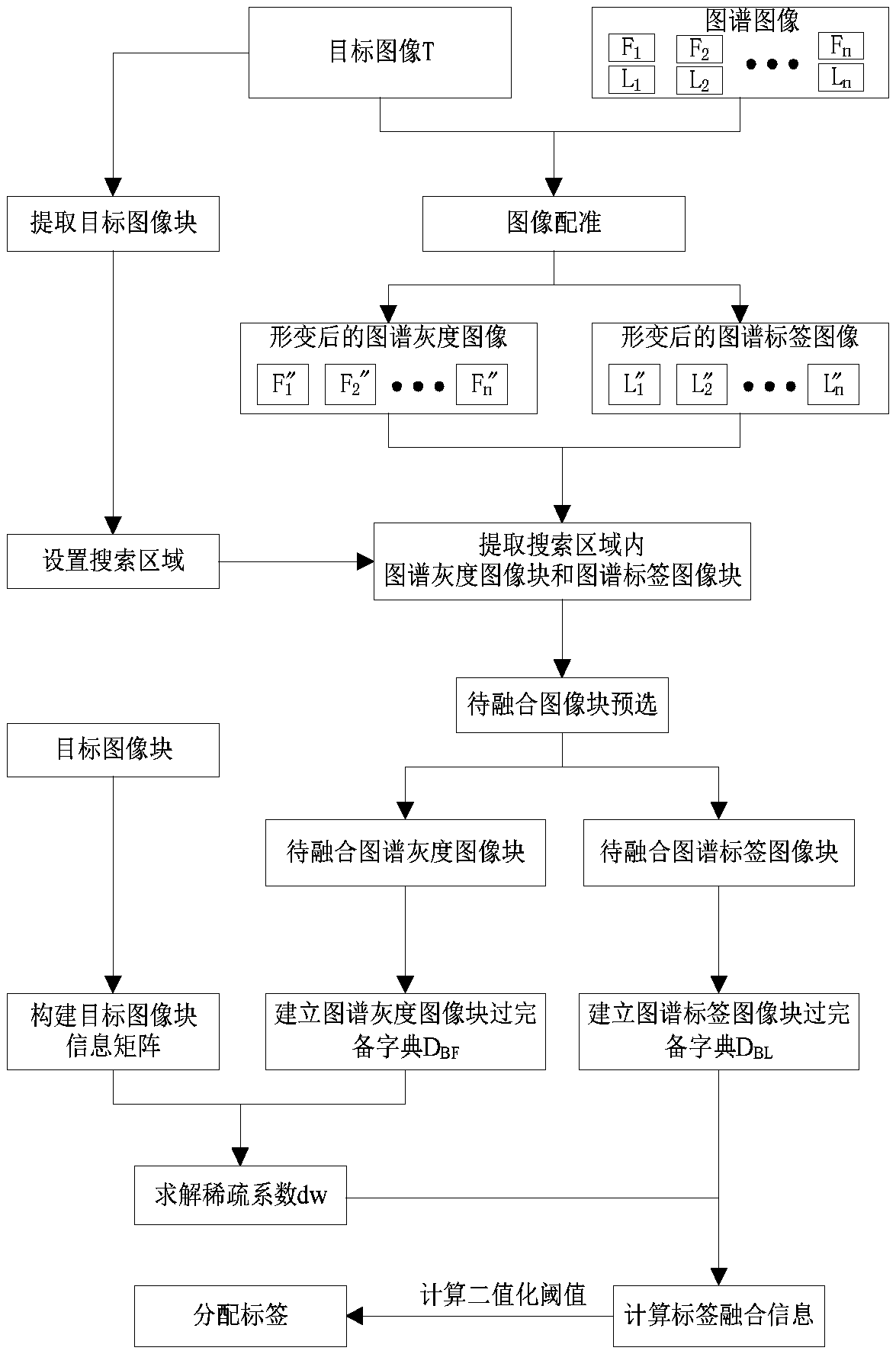
[0054] Such as figure 1 As shown, the specific implementation process of a medical image automatic segmentation method based on multi-atlas label fusion described in this embodiment is:

[0055] Step 1. Atlas registration;

[0056] Given a human brain atlas containing white matter and gray matter markers, the atlas contains N grayscale images F i (i=1,2...N) and the spectrum grayscale image F i The corresponding atlas label image L i (i=1,2…N), the L i for manual from F i The image of the target tissue is marked in , and the target image T is compared with each F i for registration.

[0057] The specific process of map registration is as follows:

[0058] First to T and F i (i=1,2...N) to normalize the pixel values ​​to reduce the error caused by t...


PUM


Abstract

The invention discloses a medical image automatic segmentation method based on multi-spectral label fusion, in particular comprising the following steps: firstly, mapping the spectral gray image and the spectral label image to a target image through spectral registration; Then,selecting the map image blocks with high similarity to the target image blocks through the tag search area setting and thepreselection of the image blocks to be fused, so as to avoid the influence of the dissimilar map image blocks on the fusion result; Finally, acquiring the final image segmentation result by establishing a sparse representation model and target pixel point label assignment step, and dynamically setting the binarization threshold is according to the obtained fusion information in the target pixel point label assignment process, in order to reduce the number of pixel points with wrong label assignment and improve the segmentation accuracy. A method accord to that invention can obtain repeatableresults and is not affect by human subjective factors.

Description

technical field [0001] The invention relates to the technical field of image segmentation, in particular to an automatic medical image segmentation method based on multi-atlas label fusion. Background technique [0002] Medical image segmentation is a key step in the field of medical image processing and analysis. Its purpose is to segment normal tissue, diseased tissue, or areas with some special meaning in the image for subsequent operations such as 3D reconstruction and quantitative analysis. Medical image segmentation can provide a reliable basis for clinical diagnosis and pathology research, and assist doctors to make more accurate diagnoses. [0003] There are three kinds of medical image segmentation methods: manual segmentation, semi-automatic segmentation and automatic segmentation. Manual segmentation and semi-automatic segmentation have certain requirements for people's work experience and have strong subjectivity. Therefore, it is particularly important to study ...

Claims


Application Information

Patent Timeline
IPC IPC(8): G06T7/00G06T7/11G06T7/136G06T7/30
CPCG06T7/0012G06T7/11G06T7/136G06T7/30G06T2207/10088G06T2207/30016
Inventor 王沫楠李鹏程
Owner HARBIN UNIV OF SCI & TECH
Learn More