
Method for focusing camera image in digital slice scanner

A technology of digital slicing and focusing method, applied in the field of image processing, can solve the problems of image out of focus, many fields of view, lost focus, etc., to achieve fast focusing speed and solve the effect of image out of focus

Active Publication Date: 2019-11-05
山东志盈医学科技有限公司
View PDF3 Cites 5 Cited by
  • Summary
  • Abstract
  • Description
  • Claims
  • Application Information

AI Technical Summary

Problems solved by technology

In the process of finding the focus, if the judgment is made according to the entire moving distance of the scanner's Z axis, the field of view to be judged is too much and the speed is too slow; if the search is made according to a selected moving range, there is a possibility of losing the focus, resulting in image out of focus

Method used


Image

  • Method for focusing camera image in digital slice scanner
  • Method for focusing camera image in digital slice scanner
  • Method for focusing camera image in digital slice scanner

Examples


Embodiment Construction

[0046] Below in conjunction with accompanying drawing and embodiment the present invention will be further described:

[0047] In order to clearly illustrate the technical features of this solution, the present invention will be described in detail below through specific implementation modes and in conjunction with the accompanying drawings. The following disclosure provides many different embodiments or examples for implementing different structures of the present invention. To simplify the disclosure of the present invention, components and arrangements of specific examples are described below. Furthermore, the present invention may repeat reference numerals and / or letters in different instances. This repetition is for the purpose of simplicity and clarity and does not in itself indicate a relationship between the various embodiments and / or arrangements discussed. It should be noted that components illustrated in the figures are not necessarily drawn to scale. Description...


PUM


Abstract

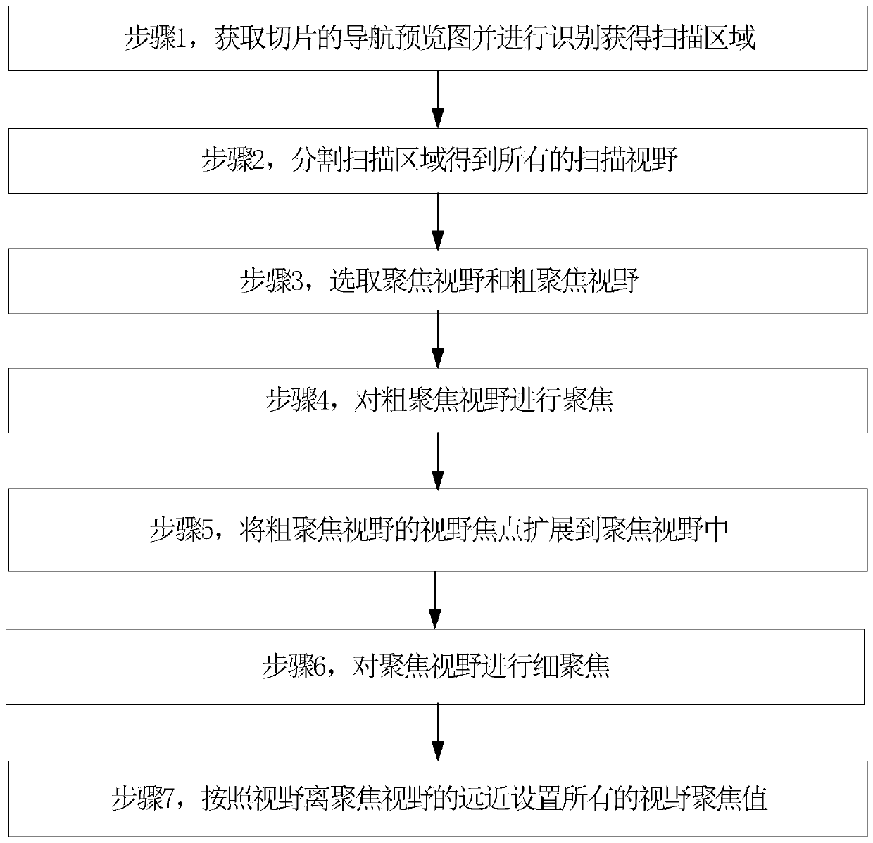
The invention discloses a method for focusing a camera image in a digital slice scanner. The method comprises the following steps: 1, obtaining a navigation preview image of a slice and identifying and obtaining a scanning area; 2, dividing all scanning visual fields in the scanning area; 3, selecting a focussed visual field and a coarse focusing visual field; 4, focusing the coarse focusing visual field; 5, expanding the field focus of the coarse focusing field into the focusing field; 6, finely focusing the focusing visual field; 7, setting focus values of all visual field according to the distances from the visual fields to the focused visual field. The method has high stepping focusing speed, an image focus can be accurately found, the method is used for a full-automatic digital pathological scanner, and the problem of image defocusing in an existing camera image focusing technology is solved.

Description

technical field [0001] The invention relates to a camera image focusing method in a digital slice scanner, belonging to the technical field of image processing. Background technique [0002] Pathological examination is a pathological and morphological method used to examine pathological changes in body organs, tissues or cells. In order to explore the disease process of organs, tissues or cells, digital pathological slide scanners can be used to examine the pathological morphological examination of their lesions, and discuss the causes, pathogenesis, and development of lesions. process, and finally make a pathological diagnosis and pathological morphological examination. [0003] The digital pathology slide scanner combines the micro-optical magnification system and computer technology to scan and collect images of traditional pathological glass slices one by one, and splicing and merging each pair of images to form a high-resolution, full-information digital pathology sli...

Claims


Application Information

Patent Timeline
Patent Type & Authority Applications(China)
IPC IPC(8): G01N21/01G01N21/84G02B7/38H04N5/232
CPCG01N21/01G01N21/84G02B7/38H04N23/675H04N23/67H04N23/632
Inventor 耿世超
Owner 山东志盈医学科技有限公司
Learn More