Image feature extraction method and device, tumor recognition system and storage medium
An image feature extraction and tumor technology, applied in the field of medical image processing, can solve the problem that the image features of tumor identification cannot be extracted
- Summary
- Abstract
- Description
- Claims
- Application Information
AI Technical Summary
Problems solved by technology
Method used
Image
Examples
Embodiment 1
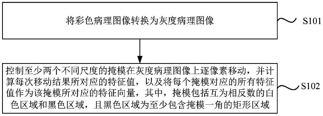
[0049] figure 1 It is a flow chart of the image feature extraction method provided by Embodiment 1 of the present invention. The technical solution of this embodiment is suitable for automatic tumor recognition based on pathological images. The method can be executed by the image feature extraction device provided in the embodiment of the present invention, and the device can be implemented in the form of software and / or hardware, and configured to be applied in a processor. The method specifically includes the following steps:
[0050] S101. Acquire grayscale pathological images of pathological slices.
[0051]Wherein, the images of the physiological slices collected by the medical microscope are usually color pathological images. If the resolutions of the collected color pathological images are the same, the collected color pathological images are directly converted into grayscale pathological images. If the resolutions of the collected color pathological images are diff...
Embodiment 2
[0070] Figure 4 It is a structural block diagram of an image feature extraction device provided in Embodiment 2 of the present invention. The device is used to execute the image feature extraction method provided in any of the above embodiments, and the device may be implemented by software or hardware. The unit includes:
[0071] Acquisition module 11, for obtaining the gray-scale pathological image of pathological slice;
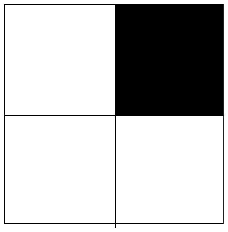
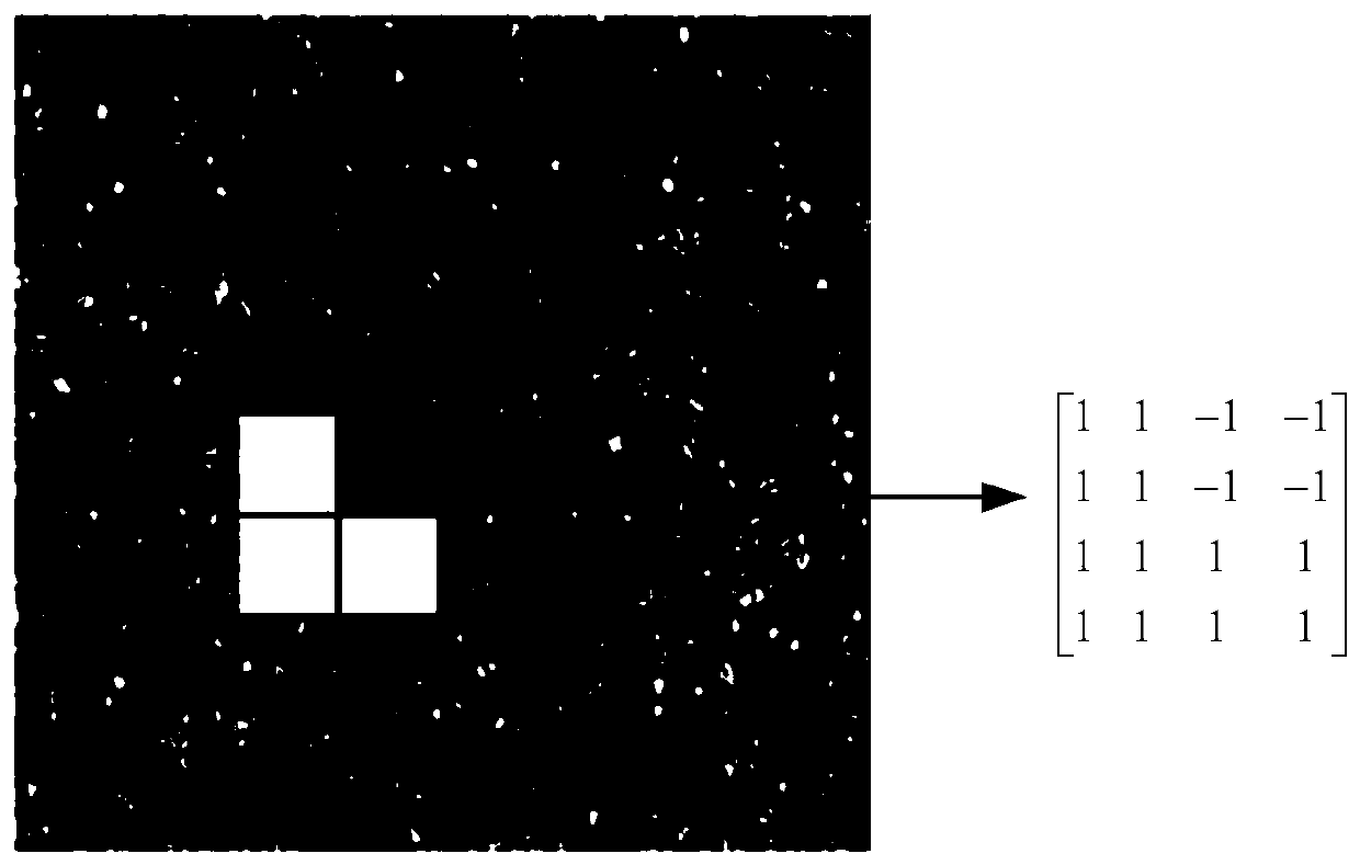
[0072] The eigenvector module 12 is used to control at least two masks of different scales to move pixel by pixel on the grayscale pathological image, and calculate the eigenvalues corresponding to each movement result, and stitch all the eigenvalues corresponding to each mask A feature vector used to represent the structural features of the grayscale pathological image is formed together, wherein the mask includes a white area and a black area which are mutually opposite numbers, and the black area is a rectangular area including at least one corne...
Embodiment 3
[0083] Figure 5 Schematic diagram of the structure of the tumor recognition system provided in Example 3 of the present invention, such as Figure 5 As shown, the system includes a processor 201 and a memory 202; the number of processors 201 in the system can be one or more, Figure 5 Take a processor 201 as an example; the processor 201 and the memory 202 in the system can be connected by bus or other methods, Figure 5 Take connection via bus as an example.
[0084] The memory 202, as a computer-readable storage medium, can be used to store software programs, computer-executable programs and modules. The processor 201 executes various functional applications and data processing of the device by running the software programs, instructions and modules stored in the memory 202, that is, implements the following methods:
[0085] Obtain the gray-scale pathological image of the pathological slice; control at least two masks of different scales to move pixel by pixel on the gr...
PUM
 Abstract
Description
Claims
Application Information
 - R&D
- Intellectual Property
- Life Sciences
- Materials
- Tech Scout
- Unparalleled Data Quality
- Higher Quality Content
- 60% Fewer Hallucinations
Browse by: Latest US Patents, China's latest patents, Technical Efficacy Thesaurus, Application Domain, Technology Topic, Popular Technical Reports.
© 2025 PatSnap. All rights reserved.Legal|Privacy policy|Modern Slavery Act Transparency Statement|Sitemap|About US| Contact US: help@patsnap.com


