
Medical image processing method and device, electronic equipment and computer storage medium

A medical image and processing method technology, applied in the field of image processing, can solve problems such as unsatisfactory detection effect, poor algorithm performance, and excessive algorithm calculation

Active Publication Date: 2020-02-18
TENCENT TECH (SHENZHEN) CO LTD
View PDF10 Cites 21 Cited by
  • Summary
  • Abstract
  • Description
  • Claims
  • Application Information

AI Technical Summary

Problems solved by technology

However, due to the large amount of data of 3D medical images, when processing the 3D medical images based on the 2D neural network model, the detection effect will be unsatisfactory. When processing the 3D medical images based on the 3D neural network model, due to the large amount of data , and the number of parameters of the model is also large, resulting in an excessive amount of calculation of the algorithm and affecting the performance of the model
Therefore, the algorithms in the prior art have poor performance and cannot meet the processing requirements for 3D medical images.

Method used


Image

  • Medical image processing method and device, electronic equipment and computer storage medium
  • Medical image processing method and device, electronic equipment and computer storage medium
  • Medical image processing method and device, electronic equipment and computer storage medium

Examples


Embodiment Construction

[0103] In order to make the purpose, features and advantages of the present invention more obvious and understandable, the technical solutions in the embodiments of the present invention will be clearly and completely described below in conjunction with the accompanying drawings in the embodiments of the present invention. Obviously, the described The embodiments are only some of the embodiments of the present invention, but not all of them. Based on the embodiments of the present invention, all other embodiments obtained by those skilled in the art without making creative efforts belong to the protection scope of the present invention.

[0104] Embodiments of the present invention are described in detail below, examples of which are shown in the drawings, wherein the same or similar reference numerals denote the same or similar elements or elements having the same or similar functions throughout. The embodiments described below by referring to the figures are exemplary only f...


PUM


Abstract

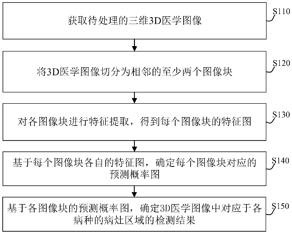
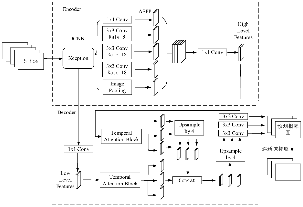
The embodiment of the invention provides a medical image processing method and device, electronic equipment and a computer storage medium. The method can comprise the steps of obtaining a to-be-processed 3D medical image; segmenting the 3D medical image into at least two adjacent image blocks; performing feature extraction on each image block to obtain a feature map of each image block; determining a prediction probability graph corresponding to each image block based on the respective feature graph of each image block; and based on the prediction probability graph of each image block, determining a detection result of a lesion area corresponding to each disease in the 3D medical image. Through the scheme of the invention, the detection result corresponding to the same disease in the 3D medical image can be accurately determined based on the image blocks and the relevance among the image blocks.

Description

technical field [0001] The present invention relates to the technical field of image processing, in particular, the present invention relates to a medical image processing method, device, electronic equipment and computer storage medium. Background technique [0002] In order to detect the location of the lesion in the 3D medical image, the existing medical image processing method is: based on the 2D or 3D neural network model, the 3D medical image is detected to determine whether there is a lesion in the medical image, and whether the lesion is in the image s position. However, due to the large amount of data of 3D medical images, when processing the 3D medical images based on the 2D neural network model, the detection effect will be unsatisfactory. When processing the 3D medical images based on the 3D neural network model, due to the large amount of data , and the number of parameters of the model is also large, which leads to an excessive amount of calculation of the alg...

Claims


Application Information

Patent Timeline
Patent Type & Authority Applications(China)
IPC IPC(8): G06T7/155G16H30/20
CPCG06T7/155G16H30/20G06T2207/10012Y02A90/10
Inventor 曹世磊刘小彤马锴伍健荣朱艳春李仁陈景亮杨昊臻常佳郑冶枫
Owner TENCENT TECH (SHENZHEN) CO LTD
Learn More