Ultrasound imaging system and method

An ultrasonic imaging system and ultrasonic data technology, applied in ultrasonic/sonic/infrasonic Permian technology, ultrasonic/sonic/infrasonic image/data processing, ultrasonic/sonic/infrasonic diagnosis, etc., to save resources and improve frame rate Effect

Active Publication Date: 2020-06-02
KONINKLJIJKE PHILIPS NV
View PDF12 Cites 3 Cited by
  • Summary
  • Abstract
  • Description
  • Claims
  • Application Information

AI Technical Summary

Problems solved by technology

[0014] Additionally, this approach requires continuous data collection throughout the cardiac cycle

Method used


Image

  • Ultrasound imaging system and method
  • Ultrasound imaging system and method
  • Ultrasound imaging system and method

Examples


Embodiment Construction

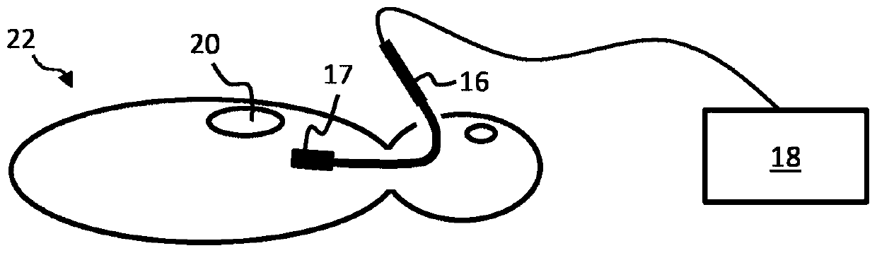
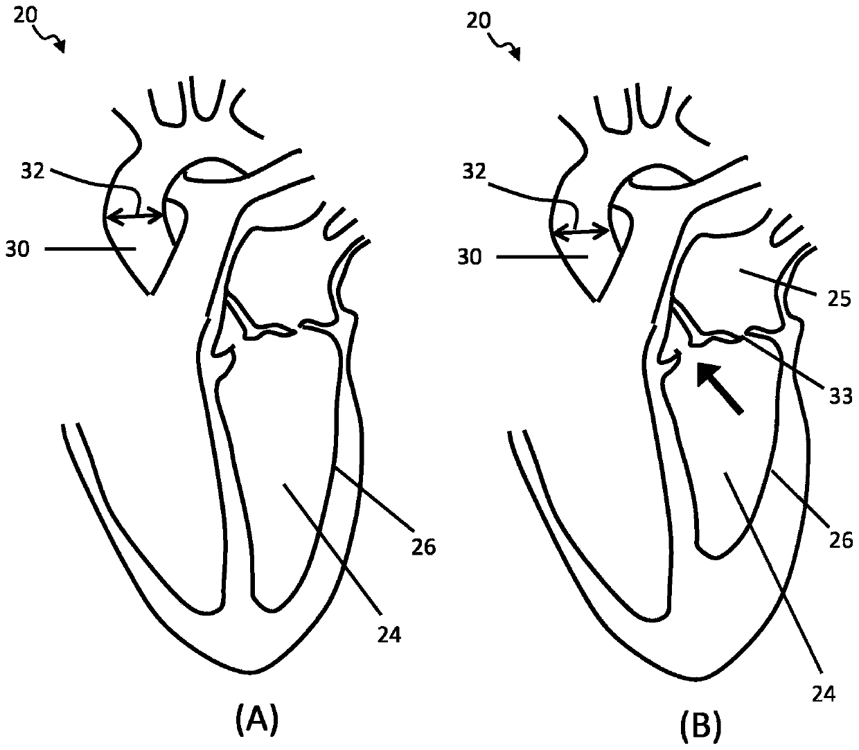
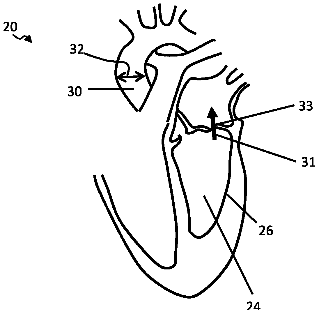
[0104] The present invention provides an ultrasound imaging system for determining stroke volume and / or cardiac output. The imaging system includes: a transducer unit for acquiring ultrasound data of a heart of a subject; and a controller. Alternatively, the imaging system may include an input for receiving acquired ultrasound data through the transducer unit rather than the unit itself. The controller is adapted to perform a two-step procedure, a first step being an initial evaluation step and a second step being an imaging step with two possible modes depending on the result of the evaluation. During the initial evaluation process, determine the presence of regurgitant ventricular flow. This is performed using Doppler processing techniques applied to the raw ultrasound data. If regurgitation is not present, stroke volume is determined using 3D ultrasound image data segmentation to identify and measure the volume of each ventricle in end-systole and end-diastole, the differ...


PUM


Abstract

An ultrasound imaging system is for determining stroke volume and / or cardiac output. The imaging system may include a transducer unit for acquiring ultrasound data of a heart of a subject (or an inputfor receiving the acquired ultrasound data), and a controller. The controller is adapted to implement a two-step procedure, the first step being an initial assessment step, and the second being an imaging step having two possible modes depending upon the outcome of the assessment. In the initial assessment procedure, it is determined whether regurgitant ventricular flow is present. This is performed using Doppler processing techniques applied to an initial ultrasound data set. If regurgitant flow does not exist, stroke volume is determined using segmentation of 3D ultrasound image data to identify and measure the volume of the left or right ventricle at each of end systole and end-diastole, the difference between them giving a measure of stroke volume. If regurgitant flow does exist, stroke volume is determined using Doppler techniques applied to ultrasound data continuously collected throughout a cardiac cycle.

Description

technical field [0001] The present invention relates to an ultrasound imaging system and method, in particular for determining stroke volume. Background technique [0002] Blood outflow from the left or right ventricle of the heart is a parameter used by clinicians in a range of diagnostic and therapeutic applications. The total blood outflow in a single cardiac cycle (single heartbeat) is called the (left ventricular) stroke volume, and the blood outflow per minute is called cardiac output. Both are key parameters used in a range of clinical applications. [0003] An important use of stroke volume is to assess IV fluid responsiveness during hemodynamic fluid intervention. [0004] During breathing, the lungs expand and contract. These volume changes result in changes in intrathoracic pressure, which is lower during inspiration than during expiration. Pressure changes have an effect on the hemodynamic function of the heart; in particular, during inspiration, intrathoraci...

Claims


Application Information

Patent Timeline
Patent Type & Authority Applications(China)
IPC IPC(8): A61B8/06A61B8/08
CPCA61B8/065A61B8/0883A61B8/12A61B8/4405A61B8/4472A61B8/469A61B8/483A61B8/486A61B8/488A61B8/5276A61B8/543A61B8/06G06T7/344A61B5/318A61B8/14A61B8/466G06T2207/10136G06T2207/20121G06T2207/30104
Inventor B·I·拉朱P·宾利F·M·韦伯J·T·萨顿T·韦克尔E·科斯滕A·鲍曼
Owner KONINKLJIJKE PHILIPS NV
Why Eureka
Try Eureka