
Mechanical ventricular pacing capture detection for a post extrasystolic potentiation (PESP) pacing therapy using at least one lead-based accelerometer

a technology of mechanical ventricular pacing and capture detection, which is applied in the field of implantable medical devices, can solve the problems of hemodynamic insufficiency, reduced the ability of the ventricle to generate contractile strength, and clinical or physician difficulties in confirming lack of therapeutic benefits, so as to improve cardiac perfusion and terminate the delivery of pesp therapy.

Inactive Publication Date: 2008-09-25
MEDTRONIC INC
View PDF8 Cites 109 Cited by
  • Summary
  • Abstract
  • Description
  • Claims
  • Application Information

AI Technical Summary

Benefits of technology

[0010]In one form, a therapy known as post-extrasystolic potentiation (PESP) therapy one (or more) pacing pulses are delivered just following the end of the refractory period of the chamber. Thus these pulses are intended to mechanically capture the chamber and provide augmented volume and flow from the chamber on successive cardiac cycles. PESP therapy delivery thus provides benefits for heart failure patients having diminished cardiac performance (i.e., cardiac insufficiency) and it has also been shown to improve cardiac perfusion following cardiac resuscitation therapy delivery (e.g., defibrillation). In this form of the invention, during a temporal window following a mechanically- and / or electrically-sensed cardiac event (e.g., ventricular contraction) and while the PESP therapy delivery is occurring signals from the motion sensor are monitored to determine if an intended (second) mechanical capture event is detected for a given cardiac cycle. If not, the PESP therapy delivery parameters can be adjusted, a mode switch can occur to a different pacing mode, or the PESP therapy delivery can be terminated.

Problems solved by technology

For example, if pacing therapy delivered to either one of the ventricles fails to mechanically capture the chamber, a clinician or physician would have difficulty confirming the lack of therapeutic benefit.
This alteration, when combined with the effects of weakened cardiac muscles, reduces the ability of the ventricle to generate contractile strength resulting in hemodynamic insufficiency.
Ventricular dyssynchrony following coronary artery bypass graft (CABG) surgery is a problem encountered relatively often, requiring post-operative temporary pacing.

Method used


Image

  • Mechanical ventricular pacing capture detection for a post extrasystolic potentiation (PESP) pacing therapy using at least one lead-based accelerometer
  • Mechanical ventricular pacing capture detection for a post extrasystolic potentiation (PESP) pacing therapy using at least one lead-based accelerometer
  • Mechanical ventricular pacing capture detection for a post extrasystolic potentiation (PESP) pacing therapy using at least one lead-based accelerometer

Examples


Embodiment Construction

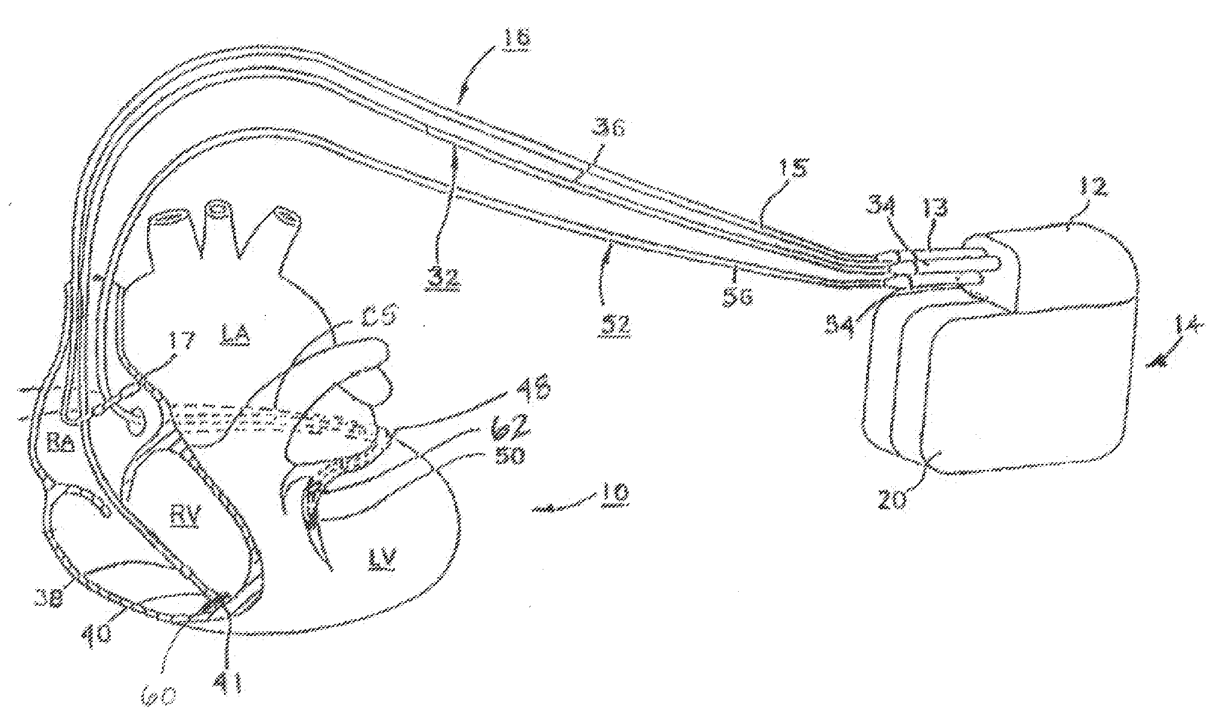
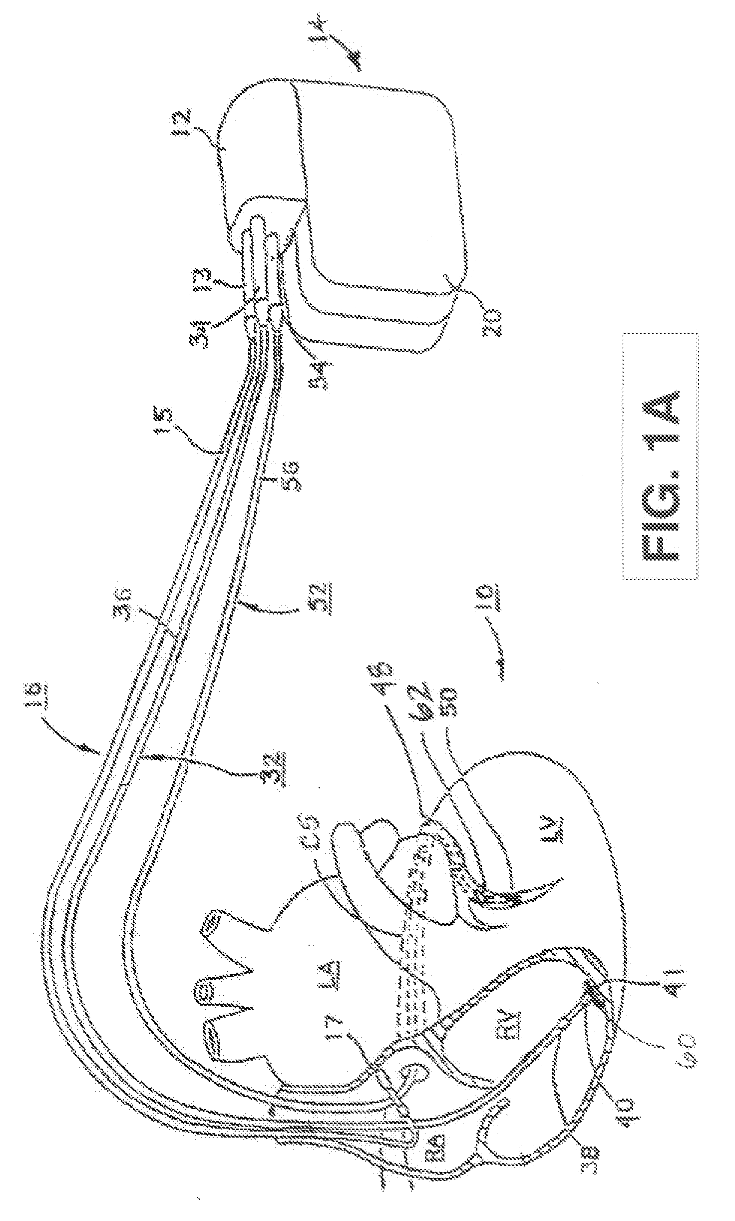
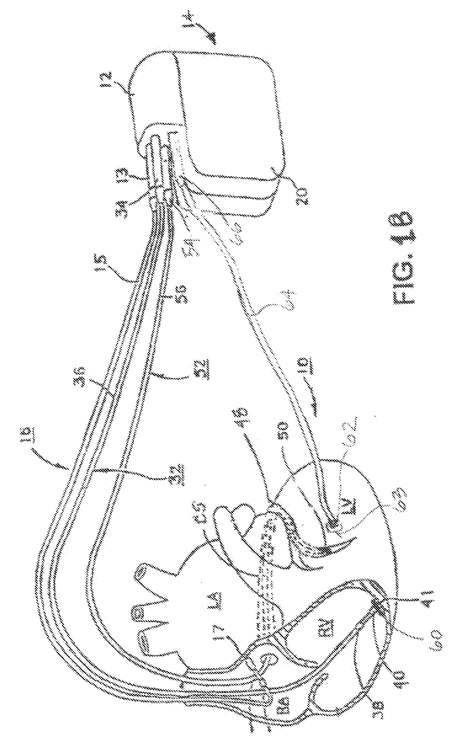
[0022]As indicated above, the present invention is directed toward providing a method and apparatus for monitoring one of more chambers of a heart for optimizing electrical pulses using mechanical parameters based on monitoring ventricular wall acceleration (e.g., LV free wall). In particular, the present invention is useful for ensuring chamber capture during pacing therapies used for treating heart failure such as PESP therapy. The present invention is also useful in ensuring capture during temporary pacing applied for treating post-operative ventricular dyssynchrony. As such, the present invention may be embodied in an implantable cardiac pacing system including a dual chamber or multichamber pacemaker and associated set of leads. Alternatively, the present invention may be embodied in a temporary pacing system including an external pacing device with associated temporary pacing leads.

[0023]FIG. 1A depicts an exemplary implantable, multi-chamber cardiac pacemaker 14 in which the ...


PUM


Abstract

A system and method for monitoring at least one chamber of a heart (e.g., a left ventricular chamber) during delivery of extrasystolic stimulation to determine if the desired extra-systole (i.e., ventricular mechanical capture following refractory period expiration) occurs. The system includes an implantable or external cardiac stimulation device in association with a set of leads such as epicardial, endocardial, and / or coronary sinus leads equipped with motion sensor(s). The device receives and processes acceleration sensor signals to determine a signal characteristic indicative of chamber capture resulting from one or more pacing stimulus delivered closely following expiration of the refractory period. A threshold optimization method optionally evaluates capture and at least one of: runs an iterative routine to establish or re-establish chamber capture for the PESP therapy, sets a logical flag relating to chamber capture status and stores parameter(s) relating to successful chamber capture for one or more subsequent cardiac cycles.

Description

STATEMENT OF INCORPORATION BY REFERENCE[0001]The present disclosure incorporates U.S. Pat. Nos. 7,142,929 entitled, “RECONFIGURABLE, FAULT TOLERANT MULTIPLE-ELECTRODE CARDIAC LEAD SYSTEMS” and 7,142,916 entitled, “CARDIAC PACING MODALITY HAVING IMPROVED BLANKING, TIMING, AND THERAPY DELIVERY METHODS FOR EXTRASYSTOLIC STIMULATION PACING THERAPY” as if fully set forth herein.FIELD OF THE INVENTION[0002]The present invention relates generally to implantable medical devices for monitoring or treating cardiac dysfunction by altering mechanical contractile function, and more particularly to devices and methods that requires the optimization of more than one electrical pulse for evoking the desired mechanical response during ventricular pacing, whether such pacing involves ventricles (i.e., bi-ventricular) or a single chamber delivery or combinations thereof (e.g., pacing pulses between ventricular configurations for contractility modulation, refractory period stimulation, stroke volume au...

Claims


Application Information

Patent Timeline
IPC IPC(8): A61N1/365A61B5/02
CPCA61N1/3712A61N1/3627
Inventor CHINCHOY, EDWARDHESS, MICHAEL F.
Owner MEDTRONIC INC
Learn More