Colposcope image recognition method for detecting cervical lesions

A technology of image recognition and cervical lesions, applied in the field of image recognition, can solve the problems of high cost, high incidence and mortality of cervical cancer, and high complexity of the overall process of cervical cancer screening, so as to improve precision and accuracy and enhance image Identify segmentation effects, effects that improve image quality

Active Publication Date: 2020-07-14
福建省妇幼保健院
View PDF5 Cites 2 Cited by
  • Summary
  • Abstract
  • Description
  • Claims
  • Application Information

AI Technical Summary

Benefits of technology

This invention improves how images are analyzed for better understanding compared with previous methods that only focused on specific areas or characteristics within them. It uses techniques like this to divide an entire picture into smaller parts called “pixels” instead of just one large area. These small regions represent different attributes such as color texture, shape, etc., but they still have their own unique properties when combined together. By performing these operations at various layers inside the pictures, we get clearer insights about what makes up our scene being captured differently from other scenes due to factors like light conditions. Overall, it provides improved results through enhanced analysis capabilities and increased resolution ability.

Problems solved by technology

This patents discusses various methods used during medical examinations on cervicitis (C), with different technical effects being discussed depending upon factors like patient risk or cost consideration. However, current techniques require complicated procedures such as histopathology, making it difficult to apply them effectively across large geographic regions where resources may lack.

Method used


Image

  • Colposcope image recognition method for detecting cervical lesions
  • Colposcope image recognition method for detecting cervical lesions
  • Colposcope image recognition method for detecting cervical lesions

Examples


Embodiment Construction

[0076] The following will clearly and completely describe the technical solutions in the embodiments of the present invention with reference to the accompanying drawings in the embodiments of the present invention. Obviously, the described embodiments are only some, not all, embodiments of the present invention. Based on the embodiments of the present invention, all other embodiments obtained by persons of ordinary skill in the art without creative efforts fall within the protection scope of the present invention.

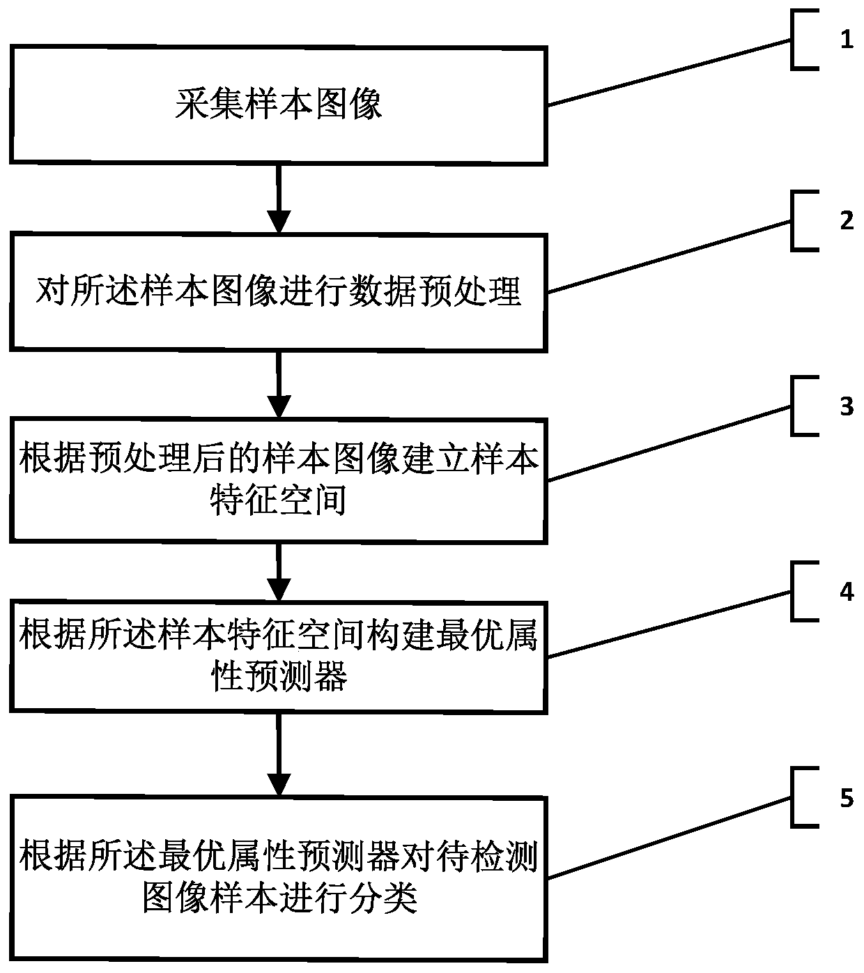
[0077] Such as figure 1 , is a flow chart of the steps of the colposcope image recognition method for detecting cervical lesions in the present invention, comprising the following steps:

[0078] Step 1. Collect sample images; the sample images mentioned in this application refer to a certain number of colposcopy images used to learn and build a training classification model. The specific acquisition method is: select a high frame rate, high resolution cervical im...


PUM


Abstract

The invention relates to the technical field of image recognition, in particular to a colposcope image recognition method for detecting cervical lesions. The method comprises the following steps: 1, collecting a sample image; 2, carrying out data preprocessing on the sample image; 3, establishing a sample feature space according to the preprocessed sample image; 4, constructing an optimal attribute predictor according to the sample feature space; and 5, classifying to-be-detected image samples according to the optimal attribute predictor. According to the technical scheme of the invention, themethod improves the attribute prediction accuracy and recall rate through adoption of the quasi-positive definite hypergraph regularization attribute learning method, can achieve the quick and accurate recognition and classification of the to-be-detected colposcope image through a learned attribute predictor, and achieves a good auxiliary detection and diagnosis effect.

Description


Claims


Application Information

Patent Timeline
Owner 福建省妇幼保健院
Why Eureka
Try Eureka