Segmentation algorithm of intracranial hemorrhage based on mu-net applied to CT images

A technology of intracranial hemorrhage and CT imaging, applied in the field of intracranial hemorrhage lesion segmentation algorithm, can solve the problems of poor U-Net segmentation performance, achieve the effect of improving segmentation performance, increasing receptive field, and solving semantic gap

Active Publication Date: 2022-01-28
JILIN UNIV
View PDF0 Cites 0 Cited by
  • Summary
  • Abstract
  • Description
  • Claims
  • Application Information

AI Technical Summary

Problems solved by technology

[0004] In order to solve the problem of poor U-Net segmentation performance in the field of intracranial hemorrhage lesion segmentation, the present invention provides an intracranial hemorrhage lesion segmentation algorithm based on MU-Net applied to CT images

Method used


Image

  • Segmentation algorithm of intracranial hemorrhage based on mu-net applied to CT images
  • Segmentation algorithm of intracranial hemorrhage based on mu-net applied to CT images
  • Segmentation algorithm of intracranial hemorrhage based on mu-net applied to CT images

Examples


Embodiment Construction

[0052] The technical solution of the present invention will be further described below in conjunction with the accompanying drawings, but it is not limited thereto. Any modification or equivalent replacement of the technical solution of the present invention without departing from the spirit and scope of the technical solution of the present invention should be covered by the present invention. within the scope of protection.

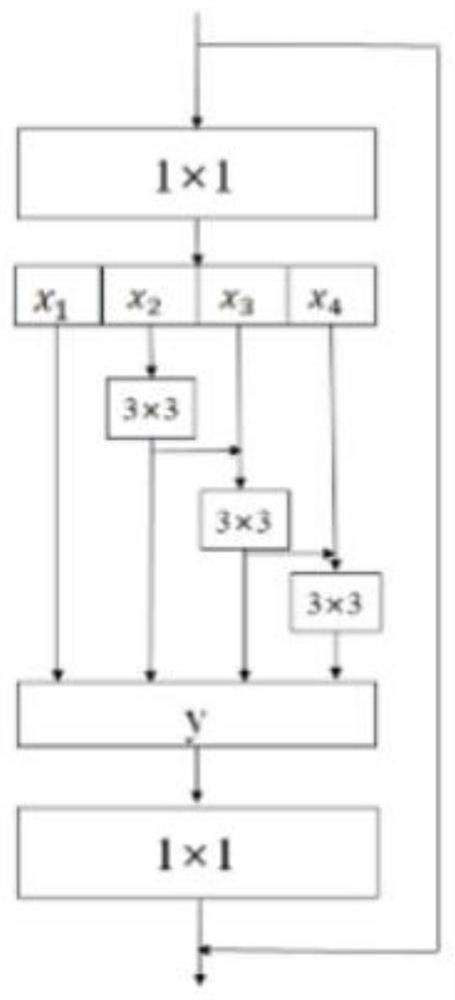
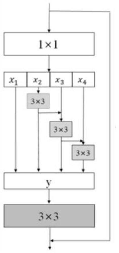
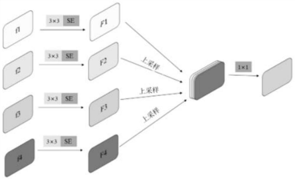
[0053]The present invention proposes a new segmentation structure MU-Net based on the U-Net, and applies it to the intracranial hemorrhage segmentation task. In the encoder module, the network module of Res2Net is introduced. Such a design can extract finer multi-scale features and increase the receptive field of feature maps. In order to reduce the semantic gap existing between the corresponding layers of the encoding layer and the decoding layer, a multi-encoding information fusion module (MIF) is proposed, which effectively compensates for the globa...


PUM


Abstract

The invention discloses an intracranial hemorrhage lesion segmentation algorithm applied to CT images based on MU‑Net. The intracranial hemorrhage lesion segmentation algorithm proposes a new segmentation structure MU‑Net based on U‑Net, and uses it Applied to the intracranial hemorrhage segmentation task. In the encoder module, the network module of Res2Net is introduced. Such a design can extract finer multi-scale features and increase the receptive field of feature maps. In order to reduce the semantic gap existing between the corresponding layers of the encoding layer and the decoding layer, a multi-encoding information fusion module (MIF) is proposed, which effectively compensates for the global information lost by the decoder through information fusion of features. Besides, in order to further reduce the semantic gap between encoder and decoder and gather multi-scale information, the present invention proposes a new decoder module (MDB).

Description

technical field [0001] The invention belongs to the field of intelligent medical image processing, and relates to an intracranial hemorrhage lesion segmentation algorithm applied to CT images based on MU-Net. Background technique [0002] Intracranial hemorrhage (ICH) is a bleeding disorder that occurs in the ventricle or brain tissue. Intracranial hemorrhage includes: intraventricular hemorrhage (IVH), parenchymal hemorrhage (IPH), subarachnoid hemorrhage (SAH), epidural hemorrhage (EDH) and subdural hematoma (SDH). Diagnosing intracranial hemorrhage is a major challenge in medicine. Determining the site of intracranial hemorrhage is key to treating the patient. The current method of diagnosing intracranial hemorrhage is a CT scan of the brain. Because tissues such as brain, blood, muscle, and bone absorb X-rays differently, CT scans produce high-contrast images that are then viewed by doctors. The research on the segmentation of intracranial hemorrhage focuses on the C...

Claims


Application Information

Patent Timeline
Patent Type & Authority Patents(China)
IPC IPC(8): G06T7/10G16H30/20G06N3/04
CPCG06T7/10G16H30/20G06T2207/10081G06T2207/20221G06T2207/30016G06T2207/20081G06N3/045
Inventor 刘萍萍宁港军石立达周求湛
Owner JILIN UNIV
Why Eureka
Try Eureka