Manipulation of meningeal lymphatic vasculature for brain and CNS tumor therapy
A technology of lymphatic and immunotherapy, applied in antineoplastic drugs, cytokines/lymphokines/interferon, angiogenin, etc.
- Summary
- Abstract
- Description
- Claims
- Application Information
AI Technical Summary
Problems solved by technology
Method used
Image
Examples
Embodiment 1
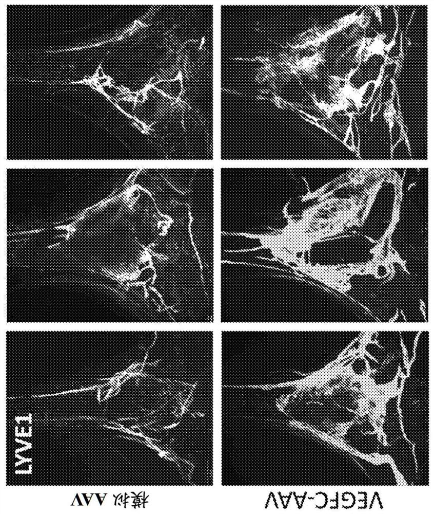
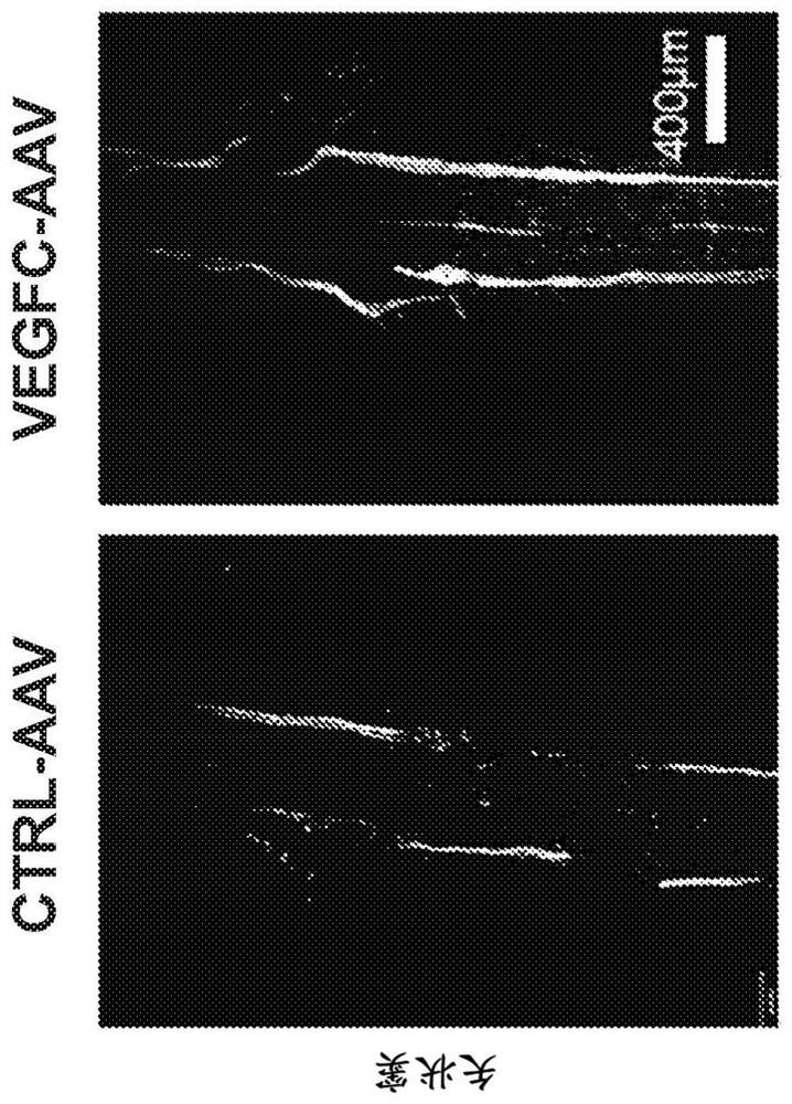
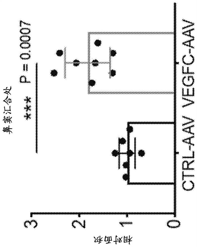
[0142] Example 1: Determination of VEGF-C Induced Lymphatic Proliferation in Dural Lymphatic Vessels
[0143] To investigate the question of how immune privilege affects tumor growth, the inventors used a model to examine tumor growth with increased meningeal lymphatic vessels.
[0144] GL261 (C57BL / 6 syngeneic GBM cell line) was transduced to express the luciferase gene (GL261-Luc) [58]. GL261 parental cells were obtained from the NIH Cancer Cell Bank. GL 261-luciferase cells were obtained from Yale Universit. GL261 and GL261-Luc cells were cultured in RPMI (10% FBS, 1% penicillin / streptomycin).
[0145] All experiments used 8-week-old in-house bred wild-type C57BL / 6 mice. C57BL / 6 mice were purchased from the National Cancer Institute and Jackson Laboratory, and subsequently bred and maintained at Yale University. All procedures were performed according to the animal experiment protocol. For tumor inoculation, intraperitoneal injections of ketamine (50 mg kg -1 ) and xy...
Embodiment 2
[0152] Example 2: Assay of VEGF-C Induction of Lymphatic Vascular Proliferation in Dural Lymphatic Vessels Manipulation of lymphatic vessels can affect the clearance of macromolecules from the brain parenchyma [34-36,50].
[0153] Mice were treated with EGFC-AAV or mock AAV as described in Example 1. A third group of wild-type mice was treated with PBS only. Go through two months.
[0154] Syngeneic mouse GBM cells (GL261-Luc) were implanted into the striatum of mice that had received AAV encoding VEGF-C, a scrambled sequence (mock AAV), or PBS (WT ). Specifically, 5,000 or 50,000 GL261-Luc cells were intracranially implanted into the striatum (the coordinates are 3 mm lateral to the bregma and 1 mm posterior). Mice were observed for signs of significant weight loss, lethargy, dorsal arch, or periorbital hemorrhage as survival endpoints. All brains were collected after death or endpoint to confirm tumor growth. 500,000 GL261 cells suspended in Matrigel (Corning) at a volu...
Embodiment 3
[0161] Example 3: Production and validation of VEGFC-mRNA
[0162] VEGFC-mRNA was custom-made from TriLink Biotechnologies, the sequence is as follows Figure 12 shown. The mRNA was produced using TriLink Biotechnologies Cap1 with full 5-methylcytosine and pseudouridine substitutions for stability. Specifically, 5-methylcytosine enhances stability and reduces deamination, while pseudouridine substitution reduces nuclease activity, reduces innate recognition and can increase translation. Cap 1 does not activate PR receptors. The mRNA is also polyadenylated (120A).
[0163] 2 μg of VEGFC-mRNA was transfected into HEK293T cells using lipofectamine. HEK293T cells were purchased from ATCC. HEK293T cells were cultured in complete DMEM (4.5 g / L glucose, 10% FBS, 1% penicillin / streptomycin). Cy5-GFP mRNA was used as a control. Cell lysates were collected 6 hours and 24 hours after transfection. Dissolve samples in RIPA buffer and boil with sample buffer for 5 min. Culture med...
PUM
 Abstract
Description
Claims
Application Information
 - R&D
- Intellectual Property
- Life Sciences
- Materials
- Tech Scout
- Unparalleled Data Quality
- Higher Quality Content
- 60% Fewer Hallucinations
Browse by: Latest US Patents, China's latest patents, Technical Efficacy Thesaurus, Application Domain, Technology Topic, Popular Technical Reports.
© 2025 PatSnap. All rights reserved.Legal|Privacy policy|Modern Slavery Act Transparency Statement|Sitemap|About US| Contact US: help@patsnap.com


