Abdominal ultrasonic image diagnosis device
A technology of ultrasonic imaging and diagnostic equipment, which is applied in the directions of acoustic wave diagnosis, infrasonic wave diagnosis, ultrasonic/sonic wave/infrasonic wave diagnosis, etc., which can solve the problem that the detection head cannot be placed stably, the detection head does not have a buffer protection structure, and the jack detection head cannot be automatically realized. Dust-proof storage and other issues to achieve the effect of preventing bump damage and improving sealing performance
Pending Publication Date: 2021-11-12
THE AFFILIATED HOSPITAL OF QINGDAO UNIV
View PDF9 Cites 0 Cited by
- Summary
- Abstract
- Description
- Claims
- Application Information
AI Technical Summary
Problems solved by technology
[0007] In order to solve the above technical problems, the present invention provides an abdominal ultrasonic imaging diagnostic device to solve the problem that although the existing portable diagnostic device is provided with a cover plate, it cannot open and close the cover through structural improvement. The dust-proof of the socket and the dust-proof storage of the detection head are automatically realized when the board is on the board; moreover, although the existing devices are equipped with a buffer structure, the buffer structure cannot realize the frictional locking of the cover through structural improvement. ; Finally, the detection head of the existing device does not have a buffer protection structure, and the suspension of the detection head and the stable placement of the snap-in type cannot be realized through the buffer protection structure
Method used
View moreImage
Smart ImageViewing Examples
Examples
Embodiment
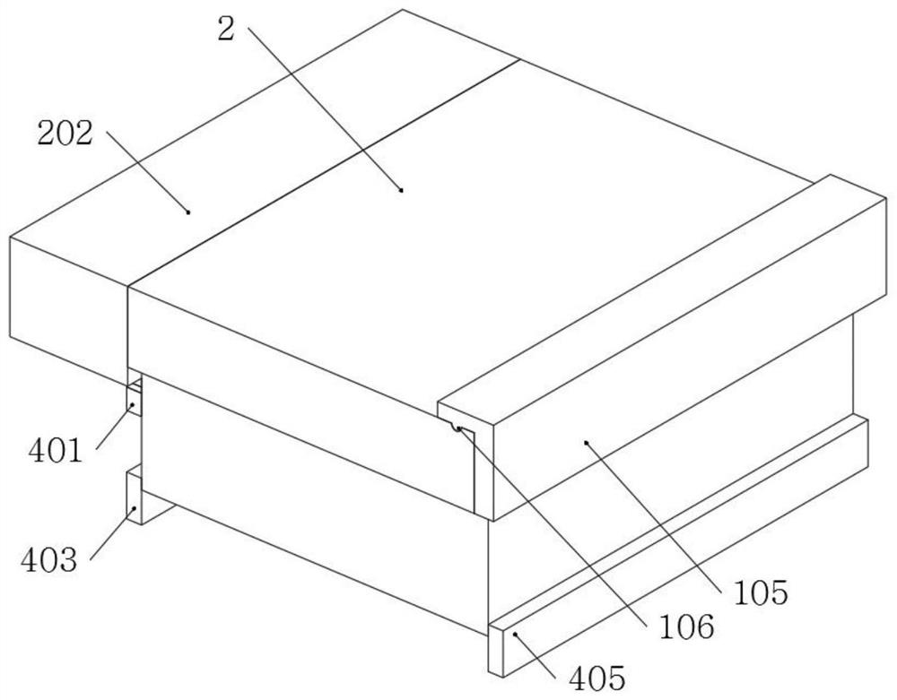
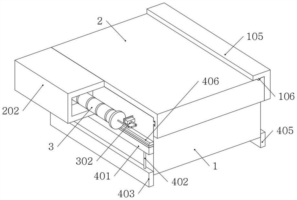
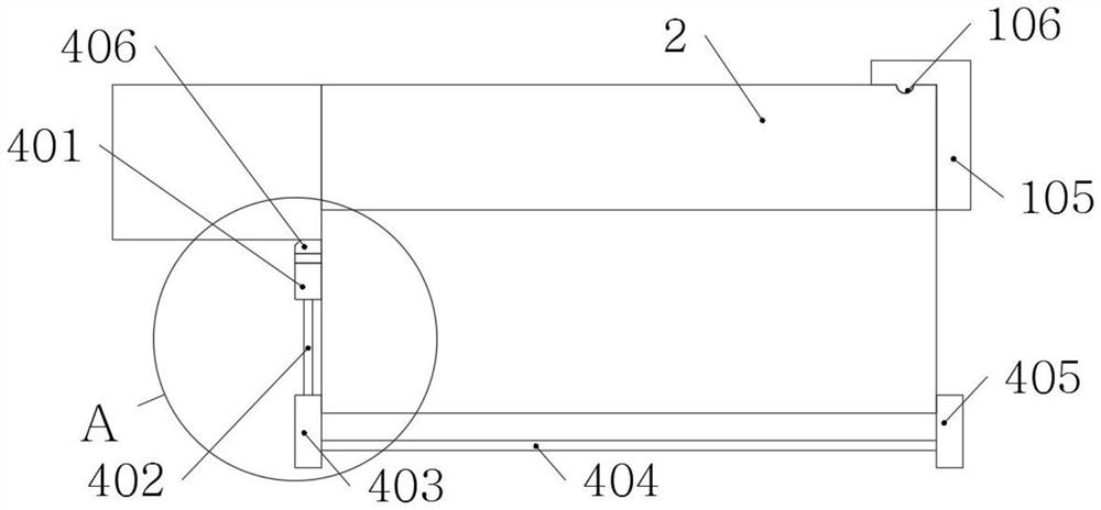
[0062] as attached figure 1 to the attached Figure 8 shown:
[0063] The present invention provides an abdominal ultrasound imaging diagnostic device, comprising a detector 1;
Login to View More PUM
 Abstract
The invention provides an abdominal ultrasonic image diagnosis device, relates to the technical field of medical equipment, and solves the problem that dust prevention of an insertion hole and dust prevention type storage of a detection head cannot be automatically realized when a cover plate is opened and closed through structural improvement, and suspension and clamping type stable placement of the detection head cannot be realized through a buffer protection structure. The abdominal ultrasonic image diagnosis device comprises a detector. The detector is of a rectangular structure. Through the arrangement of a cover plate and a buffer structure, firstly, an elastic block adheres to the top of a sliding seat, and when the cover plate is closed in a sliding mode, the top end face of the elastic block makes elastic contact with the bottom of a storage box, so that friction locking of the cover plate is achieved; and secondly, a first buffer block and a second buffer block are both of a rectangular block structure, and the bottom end face of the first buffer block and the bottom end face of the second buffer block are both lower than the bottom end face of the detector, so that the detector can be buffered through the first buffer block, the second buffer block and an elastic block when the detector falls off.
Description
technical field [0001] The invention belongs to the technical field of medical equipment, and more particularly, particularly relates to an abdominal ultrasound imaging diagnostic device. Background technique [0002] Medical imaging is biological imaging and includes diagnostic imaging, radiology, endoscopy, medical thermal imaging techniques, medical photography and microscopy. In addition, techniques including electroencephalography and magnetoencephalography, although the focus is on measurement and recording, and there is no image display, but because the data generated has localization characteristics, it can be regarded as another form of medical imaging; abdominal ultrasound; There are two types of diagnostic devices: indoor use and outdoor use. [0003] For example, the publication number: CN211049415U provides an abdominal imaging diagnostic device for radiology. The abdominal imaging diagnostic device for radiology includes a box body, a box cover, an ultrasonic...
Claims
Login to View More Application Information
Patent Timeline
 Patent Type & Authority Applications(China)
IPC IPC(8): A61B8/00A61B50/30
CPCA61B8/4427A61B8/4444A61B50/30A61B2050/0059A61B2050/0082
Inventor 王鹏鹏
Owner THE AFFILIATED HOSPITAL OF QINGDAO UNIV
Features
- R&D
- Intellectual Property
- Life Sciences
- Materials
- Tech Scout
- Unparalleled Data Quality
- Higher Quality Content
- 60% Fewer Hallucinations
Learn More Browse by: Latest US Patents, China's latest patents, Technical Efficacy Thesaurus, Application Domain, Technology Topic, Popular Technical Reports.
© 2025 PatSnap. All rights reserved.Legal|Privacy policy|Modern Slavery Act Transparency Statement|Sitemap|About US| Contact US: help@patsnap.com


