Method and device for identifying abnormal cell image in cervical cell image

A technology of cervical cells and abnormal cells, applied in the field of image processing, can solve the problems of abnormal cell extraction results, lack of computer-aided film reading system, etc.

Pending Publication Date: 2022-03-25
SHANGHAI PUDONG DEVELOPMENT BANK
View PDF0 Cites 6 Cited by
  • Summary
  • Abstract
  • Description
  • Claims
  • Application Information

AI Technical Summary

Problems solved by technology

[0004] However, the current traditional method of screening abnormal cells in cervical cells through computer vision technology lacks an effective computer-aided film reading system. Provide interpretability basis for the extraction results of abnormal cells

Method used


Image

  • Method and device for identifying abnormal cell image in cervical cell image
  • Method and device for identifying abnormal cell image in cervical cell image
  • Method and device for identifying abnormal cell image in cervical cell image

Examples


Embodiment Construction

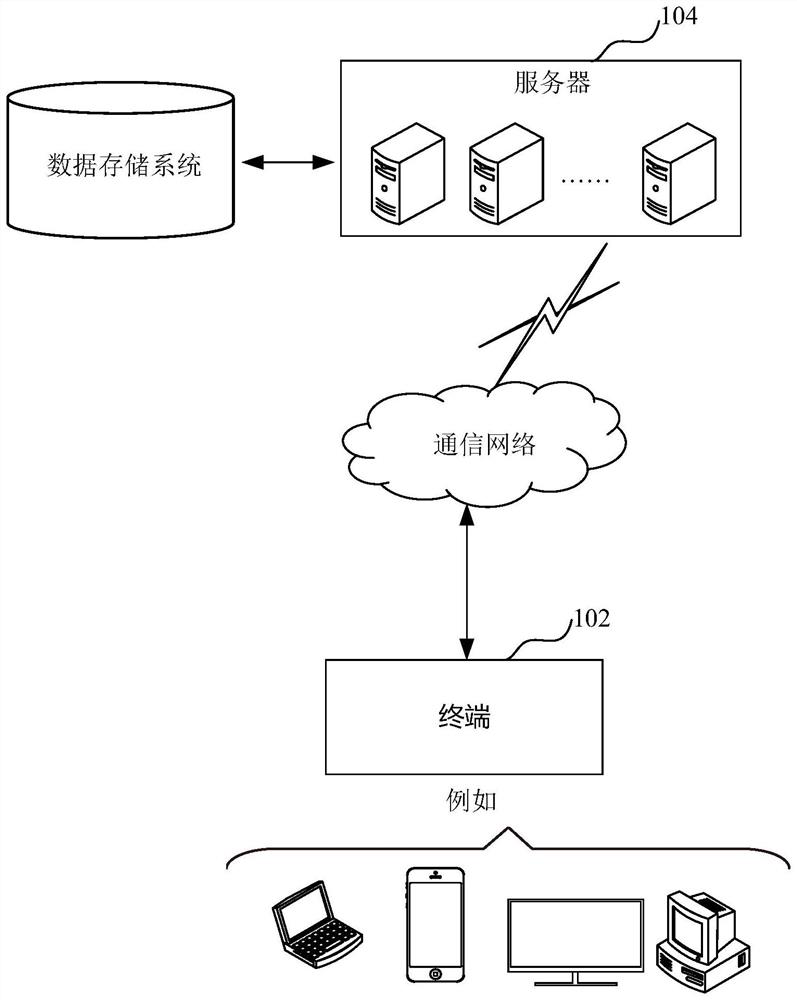
[0076] In order to make the purpose, technical solutions and advantages of the present disclosure clearer, the present disclosure will be further described in detail below in conjunction with the accompanying drawings and embodiments. It should be understood that the specific embodiments described here are only used to explain the present disclosure, not to limit the present disclosure.

[0077] It should be noted that the terms "first" and "second" in the description and claims herein and the above drawings are used to distinguish similar objects, but not necessarily used to describe a specific order or sequence. It is to be understood that the data so used are interchangeable under appropriate circumstances such that the embodiments herein described herein can be practiced in sequences other than those illustrated or described herein. Furthermore, the terms "comprising" and "having", as well as any variations thereof, are intended to cover a non-exclusive inclusion, for exam...


PUM


Abstract

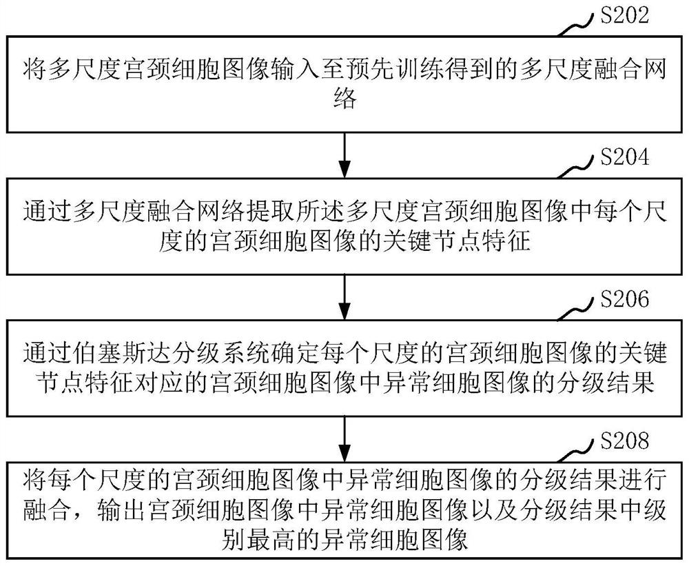
The invention relates to a method and a device for identifying an abnormal cell image in a cervical cell image. The method comprises the following steps: inputting a multi-scale cervical cell image into a pre-trained multi-scale fusion network, wherein multi-scale cervical cells comprise different sizes of the same cervical cell image; key node features of the cervical cell image of each scale in the multi-scale cervical cell images are extracted through a multi-scale fusion network; determining a grading result of an abnormal cell image in the cervical cell image corresponding to the key node feature of the cervical cell image of each scale through a Bersada grading system; and fusing the grading results of the abnormal cell images in the cervical cell images of each scale, and outputting the abnormal cell images in the cervical cell images and the abnormal cell image with the highest grade in the grading results. By adopting the method, the abnormal cell image in the cervical cell image does not need to be manually identified and determined, and the abnormal cell image can be graded.

Description

technical field [0001] The present disclosure relates to the technical field of image processing, in particular to a method and device for identifying abnormal cell images in cervical cell images. Background technique [0002] Cervical cancer is a common gynecological malignancy, and its incidence rate ranks second among female malignancies, second only to breast cancer. Early detection and intervention of cervical cancer can effectively prevent its occurrence and development. As the main method to detect cervical cancer and precancerous lesions, cervical cytology can greatly reduce morbidity and mortality. Due to the lack of a large number of strictly trained and experienced pathological cytology doctors, it is currently difficult for my country to meet the needs of large-scale and high-level cervical cancer screening. [0003] With the development of artificial intelligence technology and its wide application in the field of medical imaging, computer-aided automatic diag...

Claims


Application Information

Patent Timeline
Patent Type & Authority Applications(China)
IPC IPC(8): G06V20/69G06V10/764G06V10/80G06V10/82G06K9/62G06N3/04G06N3/08G16H50/20
CPCG06N3/08G16H50/20G06N3/047G06N3/048G06N3/045G06F18/2433G06F18/2415G06F18/254G06F18/241
Inventor 徐敏李捷王巍张玉琦张瑞雪
Owner SHANGHAI PUDONG DEVELOPMENT BANK
Why Eureka
Try Eureka