Fine-grained cervical cell image three-stage identification method

A cervical cell and identification method technology, applied in the field of three-stage identification of fine-grained cervical cell images, to achieve the effect of complete identification process system and improved accuracy

Pending Publication Date: 2020-10-30
NANTONG UNIVERSITY
View PDF3 Cites 7 Cited by
  • Summary
  • Abstract
  • Description
  • Claims
  • Application Information

AI Technical Summary

Problems solved by technology

[0007] The technical problem to be solved by the present invention is to provide a three-stage recognition method for fine-grained cervical cell images. Starting from the practical application of cervical cell classification and recognition, explore and study the use of cost-sensitive learning methods to deal with the problem of unbalanced binary classification to remove impurities; Learn thinking methods, study the multi-classification problem of cervical cell imbalance, and improve the accuracy of classification recognition

Method used


Image

  • Fine-grained cervical cell image three-stage identification method
  • Fine-grained cervical cell image three-stage identification method

Examples


Embodiment 1

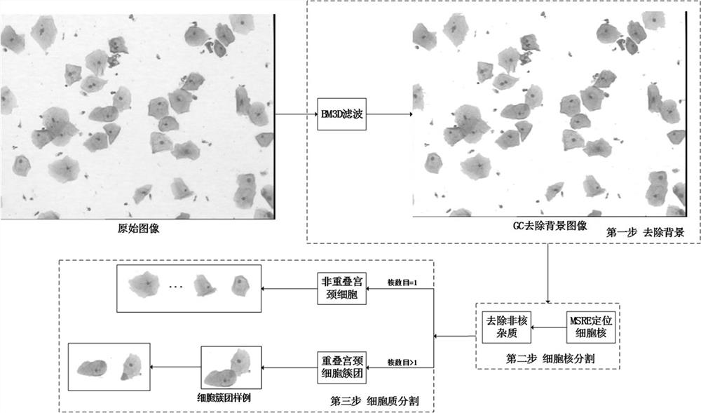
[0041] Embodiment 1: A three-stage recognition method for fine-grained cervical cell images, comprising the following steps:

[0042] (1) For the entire microscopic image of cervical cells, the three-dimensional block-matching and 3D filtering (BM3D) algorithm is used to denoise, and the speed of image preprocessing is improved by implementing BM3D based on parallel programming technology;

[0043] (2) On the filtered image, first establish a Graph Cut model to divide the image into foreground and background, and then use the MSER (Maximally Stable Extremal Region) algorithm to segment the nucleus, including the following steps:

[0044](2-1) Remove the background: on the basis of the denoised image, use the graph cut GC algorithm to remove the image background, and determine the foreground cells and cell cluster areas;

[0045] (2-2) Cell nucleus segmentation: The MSER algorithm is used to segment the cell nuclei in the foreground cells and cell cluster areas of the image.

...


PUM


Abstract

The invention provides a fine-grained cervical cell image three-stage identification method, which comprises the following steps: firstly, denoising a whole cervical cell image acquired by electronicimaging equipment to improve the image quality, removing a background to determine a foreground cell region, and segmenting out cell nucleuses and cytoplasm; secondly, extracting shape, color, textureand abstract features from the segmented cell regions, and carrying out selective fusion on the features by adopting a multi-kernel learning method so as to optimize a feature mode; and finally, removing non-cervical cell impurities according to the optimized characteristic mode, and performing multi-classification on the cervical cell images by adopting a cost-sensitive active learning method. The identification process system is complete, can extract medical features of multiple types of cervical cell images, performs selective fusion optimization on feature modes, removes impurities by using a cost-sensitive learning method, solves the problem of unbalanced dichotomy, and can effectively improve the accuracy of cervical cell classification and identification.

Description

technical field [0001] The invention belongs to the field of medical image processing and analysis of computer vision, and in particular relates to a three-stage recognition method for fine-grained cervical cell images. Background technique [0002] At present, with the progress and development of modern science and technology, great changes have taken place in the experimental methods and research directions in the field of biomedicine, and the experimental data presents a trend of "big data", involving massive amounts of clinical medical data, genomics data, and literature data. Wait. With the gradual improvement of medical infrastructure such as medical information systems, many hospitals have collected and collected more and more medical images of patients to be tested, and the same is true for cervical cell images. Accurate cervical cell image diagnosis technology can reduce the probability of misjudgment caused by the fatigue of doctors who read the film for too long,...

Claims


Application Information

Patent Timeline
Patent Type & Authority Applications(China)
IPC IPC(8): G06K9/62G06T7/00G06T7/11G06T7/13G06T7/194G06T7/90
CPCG06T7/0012G06T7/194G06T7/11G06T7/13G06T7/90G06T2207/10061G06T2207/30024G06T2207/20081G06F18/253G06F18/24
Inventor 赵理莉胡彬杨晋朝
Owner NANTONG UNIVERSITY
Why Eureka
Try Eureka