Patient specific anatomic kidney phatnom

a technology of anatomical kidney and patient, applied in the field of medical anatomical simulator of patient organs, can solve the problems of high price and limited usage possibilities, lack of different organ simulation possibilities, and excessive use of physical phantoms, and achieve realistic mechanical and imaging properties, and widen the utility possibilities of kidney phantoms.

Active Publication Date: 2018-09-25
TALLINN UNIVERSITY OF TECHNOLOGY
View PDF5 Cites 4 Cited by
  • Summary
  • Abstract
  • Description
  • Claims
  • Application Information

AI Technical Summary

Benefits of technology

[0011]Compared to the phantoms found on the market the authors have produced an affordable disposable drainage practice phantom with the possibility to practice nephrostomy which widens the kidney phantoms utility possibilities. In addition the price per phantom makes the solution according to the present invention a possible contestant on the market of renal biopsy phantoms. In general the authors of present invention believe that the patient specific phantom will be accepted by the radiology staff to use phantoms of present invention in training and in everyday practice before the surgery.
[0012]The advantages of the present patient specific anatomical kidney phantom with calyxes for drainage training in interventional radiology are obtained by manufacture anthropomorphically accurate 3D phantom organs having physiologically realistic mechanical and imaging properties. In the first step the accurate 3D shape of the organ for example of the patient kidney is created, thereafter the kidney mold was manufactured and filled with the gelatine mixture that had appropriate ultrasound properties. In another embodiment the realistic 3D shape was produced by using human abdominal CT scan which allows to manufacture very realistic organ phantom with all parts of the organ. Before casting the kidney phantom the kidney calyxes and drainage system comprising connecting pipes and reservoirs are placed in housing of the phantom according to the placement of calyxes in the patient kidney. Each reservoir has connection via connecting pipes to different parts (calyxes) of the kidney but they are not connected to each other which allow filling reservoirs during the biopsy studies with liquids of different colours and also with x-ray contrast media for training under fluoroscopy or CT guidance.

Problems solved by technology

Confidence could be raised also with patient specific organ training, which is widely used in form of surgical simulators [5], but has not yet been extensively used in physical phantoms due to high cost and limited usage of the commercial phantoms.
The market provides few phantoms that could be used for nephrostomy training which are usually with high price and limited usage possibilities.
However there is lack of possibilities to simulate different part of the organ, for example the human kidney contains number of calyxes, and it is very important to train the biopsy and / or drainage with all parts of the kidney involved.
As the kidney consist more than one objects of interest after the procedures and drainage of the organs it is impossible to evaluate if the procedure was successful in a specific part of the organ.

Method used


Image

  • Patient specific anatomic kidney phatnom
  • Patient specific anatomic kidney phatnom
  • Patient specific anatomic kidney phatnom

Examples


Embodiment Construction

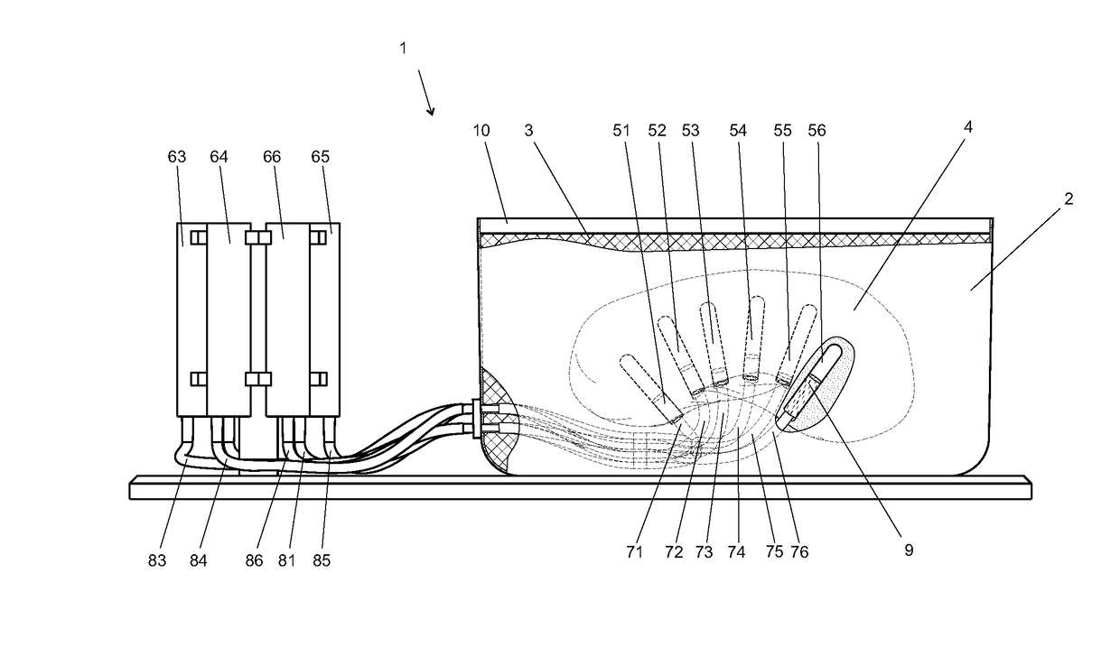
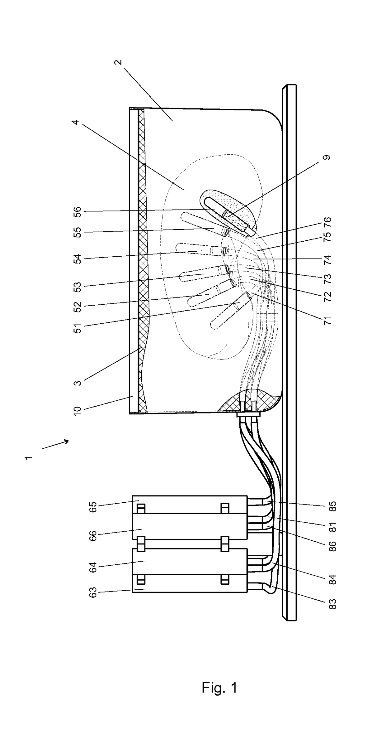
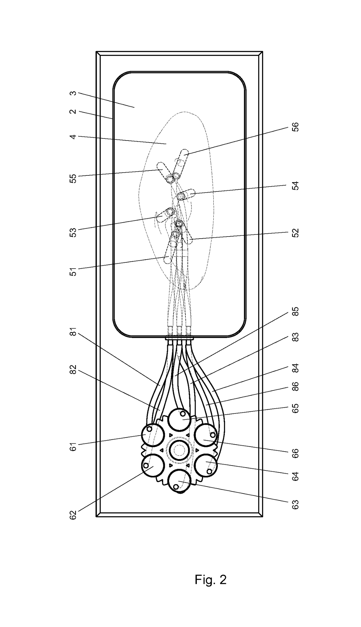
[0018]The anatomical kidney phantom simulator 1 comprises a housing 2 fixed to the base 11 and filled with the surrounding material 3 which replicates soft tissue of a patient. The housing is covered with a covering material 10 having properties corresponding to the patient skin. Into surrounding material is placed an anatomical kidney phantom 4 where said kidney phantom has a number of cavities 51, 52, 53, 54, 55, 56 simulating a parts of the kidney for drainage training in interventional radiology for example the cavities are simulating kidney calyxes. The cavities are placed in the manner which corresponds to the real placement of the kidney calyxes in the real patient kidney. Outside of the housing are outer reservoirs 61, 62, 63, 64, 65, 66 for liquids used for determining correct insertion of the catheter or for visualisation calyxes in fluoroscopy. The reservoirs can be fixed to the base 11 with connecting means or in alternative embodiment directly to the housing. As for sim...


PUM


Abstract

The invention relates to an anatonnical kidney phantom with calyces for drainage training in interventional radiology. The anatonnical kidney phantom simulator comprises a housing filled with the surrounding material and an anatomical kidney phantom placed into surrounding material. The kidney phantom has a number of cavities simulating parts of the kidney for drainage training in interventional radiology for example the cavities are simulating kidney calyces. The cavities are connected by channels and connecting pipe with outer reservoirs filled with the different colors or the x-ray opaque contrast liquids.

Description

[0001]This application is a national application of PCT-application PCT / EP2014 / 054448, filed on Mar. 7, 2014, which claims priority of the European application number EP13165254.7 filed on Apr. 24,2013, both of which are incorporated herein by reference.TECHNICAL FIELD[0002]The present invention relates to medical anatomical simulator of patient organs, and more particularly the invention relates to a patient specific anatomical kidney phantom with calyxes for drainage training in interventional radiology for example before patient surgery.BACKGROUND ART[0003]Interventional radiology has been a rapidly developing area in minimally invasive (MI) procedures due to late advances in the medical imaging [1] and rapid progress in the development of procedure equipment. MI procedures have advantages such as improved patient safety and reduced discomfort, reduced hospital stay, reduced operating room cost, etc. [2-4]. The vast amount of skills that radiology residents or other specialty res...

Claims


Application Information

Patent Timeline
Patent Type & Authority Patents(United States)
IPC IPC(8): G09B23/30G09B23/28G09B5/02
CPCG09B23/303G09B23/30G09B23/285G09B5/02
Inventor RISTOLAINEN, ASKOROSS, PEETER
Owner TALLINN UNIVERSITY OF TECHNOLOGY
Why Eureka
Try Eureka