[0008]The inventive device and method facilitates the precise preparation of both the donor and recipient bone in such a way that a large bone segment can be fit at the exact position and orientation of the preinjury tissue and in direct contact with the patient's own native bone thus restoring the articular surface and thus the smooth function of a joint.
[0009]In embodiments the key elements of the device are the articular referencing platforms and device pins. The articular referencing platforms rest on the articular surface. In such embodiments of the current invention, the articular referencing platforms are contoured to the bony surface to be prepared. Within this cutting device, there are cutting slots provided at a predetermined distance away from the articular referencing platforms and thus the articular surface of the joint. These cutting slots allow the passage of a saw blade in such a way to remove the bone segment either from the donor graft or from the recipient in such a way that both the removed donor and recipient grafts are of the same exact dimensions. When the graft is placed in the recipient site of the patient's joint, it completely restores the articular surface to the desired level with healthy articular cartilage from the donor. In order to allow for the completion of any additional angular cuts, additional guides and fixation devices are placed on the cutting guide based on the specific needs of the procedure. In one of disclosed embodiments of the invention, for the preparation of a femoral condyle, a separate attachable tower is mounted on the original cutting jig. This attachable tower facilitates the precise cutting of the diseased femoral condyle or femoral condyle graft away from the associated femoral trochlea. For example, once the femoral condylar graft has been applied into its recipient site it achieves initial stabilization to the bone through direct contact over a large bony surface and through friction between the posterior condyle, the distal condyle, and the anterior wall of the trochlea. Additional fixation is achieved with standard screws and plates as needed and as commonly practiced in the art of orthopedic surgery. The above described guides can be used alone or in combination to allow transplantation of either one or both femoral condyles. Furthermore, the guides can be utilized with a separate attached trochlear tower which rests on the two femoral condylar guides and allows for recovery of the trochlea as a single entity. Alternatively, by utilizing both condylar guides and the trochlear tower, the entire surface of the human distal femur can be transplanted. Thus the disclosed invention allows transplantation of either one or both femoral condyles independently, one femoral condyle with the femoral trochlea, the femoral trochlea in isolation, or potentially, the entire surface of the distal femur if indicated.
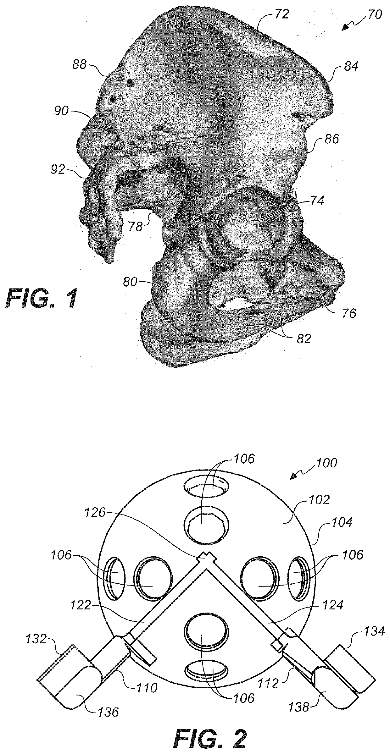
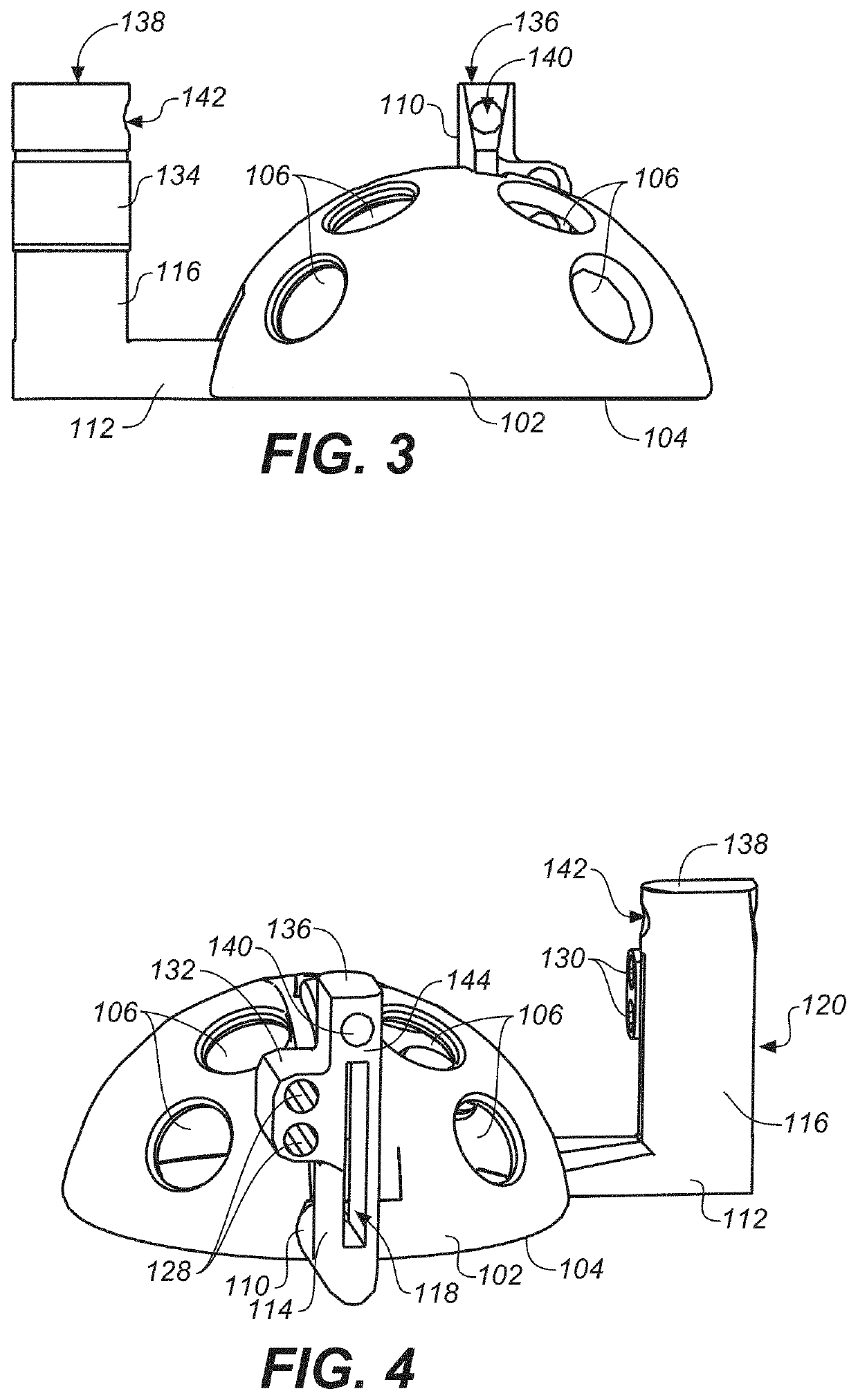
[0010]In the current document, we have expanded on the previous disclosures of devices for the preparation of the knee and disclosed additional iterations of the device, notably for the preparation of the human acetabulum or hip socket, femoral condyles, and femoral trochlea of the knee. For the human acetabulum, the majority of cases of hip arthritis start in the superior weight-bearing portion of the acetabulum. As a result, in certain cases the full resurfacing of the hip socket is not necessary. However, there is a need for a reliable method to resurface just the weight-bearing portion of the acetabulum. The current disclosure provides two methods for performing limited resurfacing of the hip socket. The first of these is a modular acetabular cutting tool that precisely contacts the articular surface of the acetabulum. This referencing guide is disposed with two cutting slots or channels, oriented radially to the base of the guide, and a set height of reference from the hemispherical articular surface of the acetabulum. The cutting slots are disposed in a radial orientation to the base of the guide and each contains a cutting channel. The radially oriented cutting slots allow the surgeon to cut orthogonally to the acetabular cartilage surface and references at an angle of between 30 and 150 degrees to one another. An axial cutting tool is then placed on the flat surface formed at the top of the radially oriented cutting slots. This axial cutting tool is disposed with a central cutting slot for placement of a saw in order to separate the bone previously cut through the radial cutting channels of the guide. This process allows for the complete separation of the acetabular graft of set thickness and essentially parallel to the articular surface at a set distance away from the cartilage. The triangular segment is removed from the donor and recipient using either reciprocating or oscillating bone saws or a cutting burr system through the three cutting slots described above (two radial and one axial).
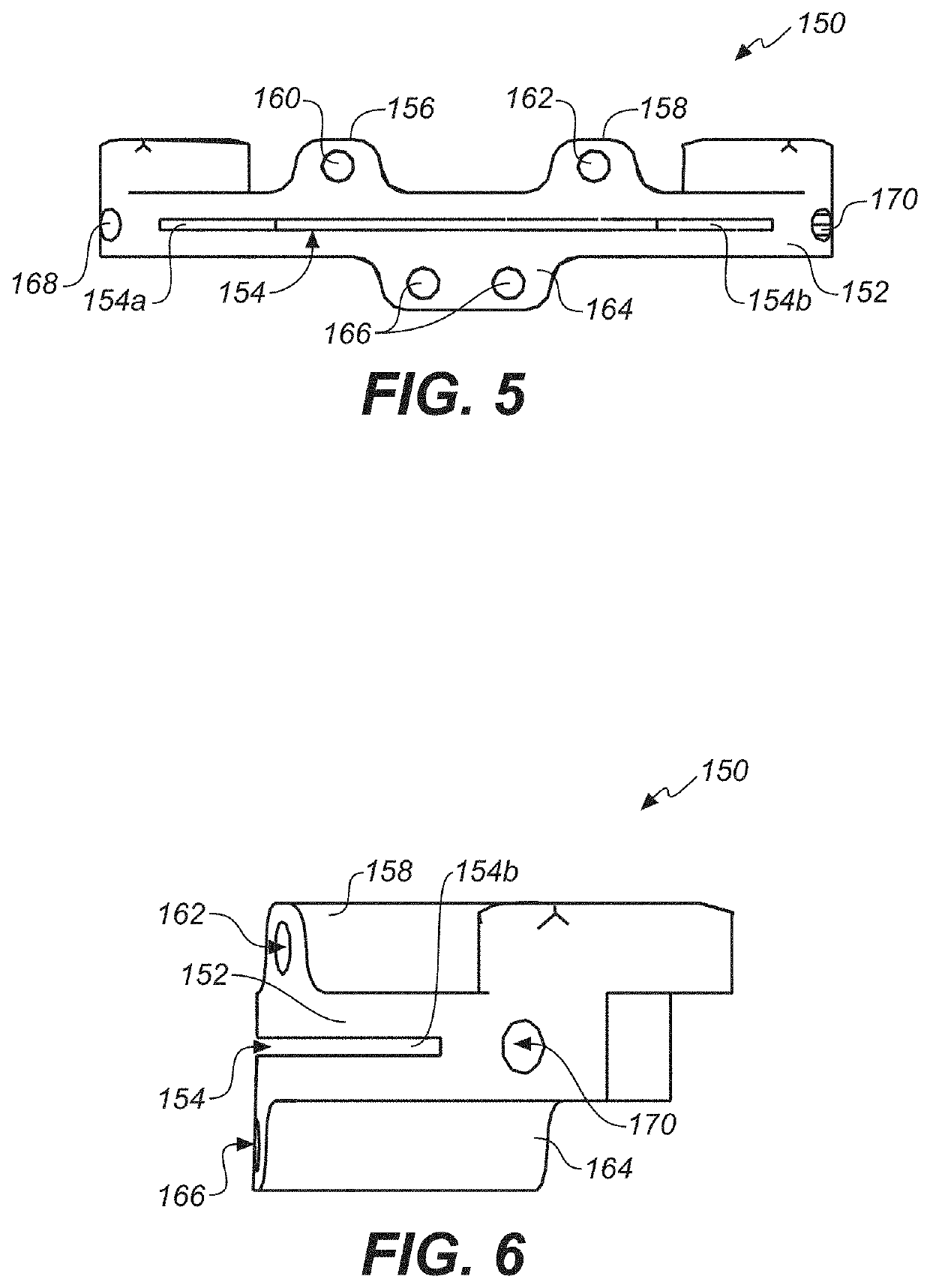
[0011]The second described cutting tool for the acetabulum, described here as the monoblock acetabular cutting guide is a unitary (one-piece) cutting block which references off the articular surface but contains the three described cutting channels within its body. It provides the advantage of easier application to the surface but the disadvantage of less visualization of the entire surface of the acetabulum.
[0012]In another embodiment, the inventive bone cutting guide is adapted for the preparation of the femoral trochlea. It shares commonalities with other embodiments based on referencing the articular surface of the joint to be resurfaced. Its unique characteristics are the use of multiple referencing pins extending through the articular side of the guide. These again serve to orient the guide at the desired distance from the articular surface of the joint to obtain a graft of set thickness and orientation. The unique qualities of this device are that it provides for the harvest of the trochlea with a triangular chevron shape that leads to increased stability after transplantation, limits the overall thickness of the graft, which is important for graft survival, and by adjustment of the reference pins allows for conversion of a malformed femoral trochlea to a trochlea with normal morphology and smooth transition to the remaining portion of the knee at the distal femur.

