
Agents for treatment of glaucomatous retinopathy and optic neuropathy
a technology of optic nerve and glaucoma, applied in the field of retinopathy and optic neuropathy, can solve the problems of not addressing the protection or treatment of cannot be predicted from the biological responses of spinal cord tissues and brain tissues, and the application cited does not address the loss of retinal ganglion cells (rgc), so as to enhance the availability or the transport of nrf2. the effect of gene products and enhancing the availability of gene products
- Summary
- Abstract
- Description
- Claims
- Application Information
AI Technical Summary
Benefits of technology
Problems solved by technology
Method used
Image

Examples
example 1
Agents Having Stimulatory Activity for Nrf2 Protein Nuclear Translocation
[0058] Vascular endothelial cells, such as bovine aortic endothelial cells (BAEC, VEC Technologies, Rensselaer, N.Y.), are used to determine those agents having stimulatory activity for Nrf2 protein nuclear translocation. For example, confluent monolayers of bovine aortic endothelial cells are exposed to candidate agents in Dulbecco's modified Eagle's medium with 1% fetal bovine serum for up to 24 hours. Cell lysates, cytosolic extracts, and nuclear extracts are prepared, and immunoblotting performed and quantified as described in Buckley, B. J., et al. (Biochem Biophys Res Commum, 307:973-979 (2003)). Agents that increase the amount of Nrf2 detected in the nuclear fraction as compared to control cells without agent are then tested for activity in a retinal ganglion cell toxicity assay as set forth in Example 2.
example 2
Protection of Rat Retinal Ganglion Cells by an Agent Having Stimulatory Activity for Nrf2 Protein Nuclear Translocation
[0059] Cultured rat neural retinal cells are combined with an agent that stimulates nuclear translocation of Nrf2 protein for 1 to 24 hours, then the combination is exposed to peroxide. Survival of the neural retinal cells as compared to a control culture without peroxide indicates that the agent provides protection from the oxidant.
[0060] Neonatal rat neural retinal cells are isolated and cultured as reported in Pang, I-H., et al, (Invest Ophthalmol Vis Sci 40:1170-1176 (1999)). “Neural retinal” refers to the retina without the retinal pigment epithelium. Thus, the culture contains a mixed population of retinal cell types. Briefly, neonatal Sprague-Dawley rats 2-5 days old are anesthetized and a 2 mm midline opening is made in the scalp just caudal to the transverse sinus. Retinal ganglion cells are selectively retrograde labeled with a fluorescent dye, Di-I, (1,...
example 3
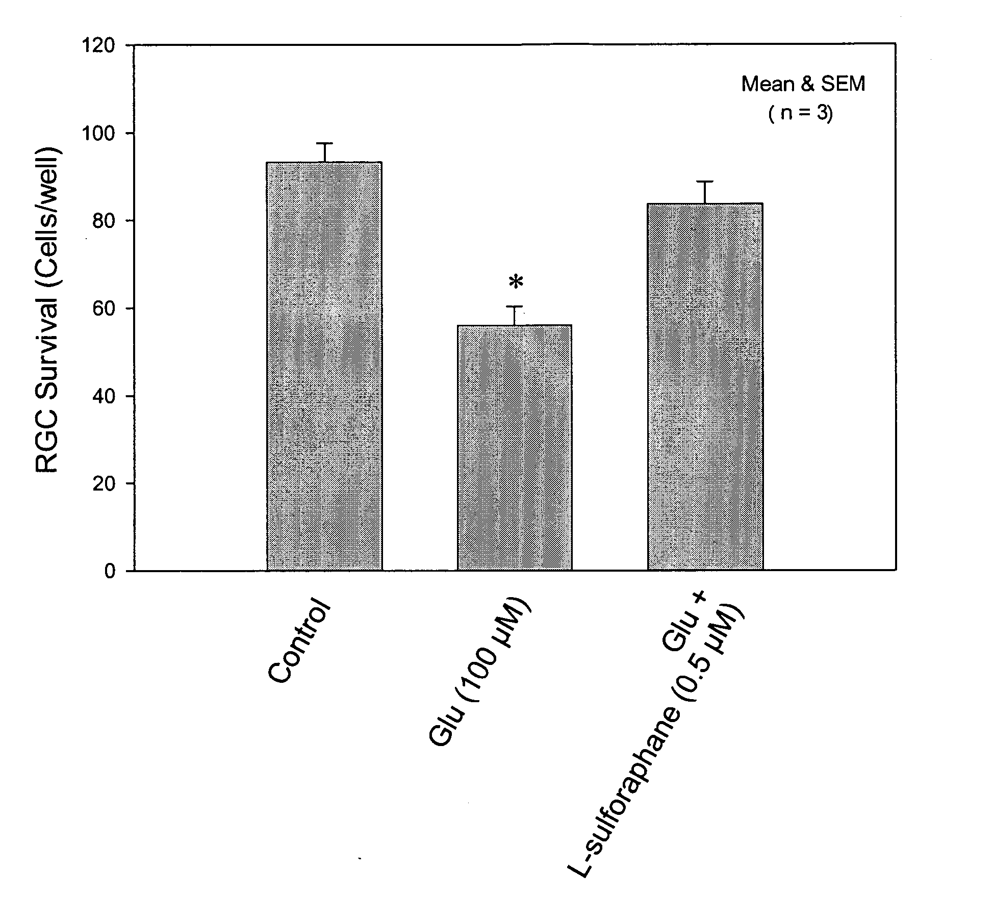
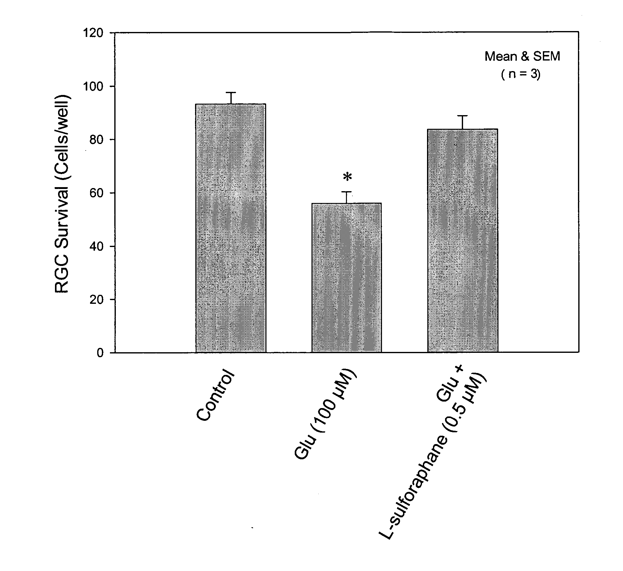
Protection of Rat Retinal Ganglion Cells from Glutamate-Induced Toxicity by Sulforaphane
[0063] Adult Sprague-Dawley rats were euthanized by CO2 asphyxiation. Their eyes were enucleated and placed in NEUROBASAL™ medium (Gibco, Gaithersburg, Md.). The retina from each eye was detached and isolated. Retinal cells were dissociated by combining up to 20 retinae with 5 mL of papain solution containing 10 mg papain, 2 mg DL-cysteine, and 2 mg bovine serum albumin in 5 ml of NEUROBASAL™ medium, for 25 min at 37° C., then washed 3 times with 5 mL RGC medium (NEUROBASAL™) medium supplemented as cited in Example 2 and with 1% fetal calf serum. Retinal pieces were triturated by passing through a fire-polished disposable pipet several times until cells were dispersed. The cell suspension was placed onto a poly-D-lysine- and laminin-coated 8-well chambered culture slide. Glutamate and glutamate with sulforaphane were added to assigned wells. The cells were then cultured at 95% air / 5% CO2 at 37° ...
PUM
 Abstract
Description
Claims
Application Information