
3D segmentation of targets in multislice image

a multi-slice image and target technology, applied in image enhancement, instruments, computing, etc., can solve the problems of unreliable segmentation of part- or non-solid nodules, and the intensity thresholding method has been shown to fail in the non-solid cas

Inactive Publication Date: 2005-09-15
SIEMENS MEDICAL SOLUTIONS USA INC
View PDF2 Cites 18 Cited by
  • Summary
  • Abstract
  • Description
  • Claims
  • Application Information

AI Technical Summary

Problems solved by technology

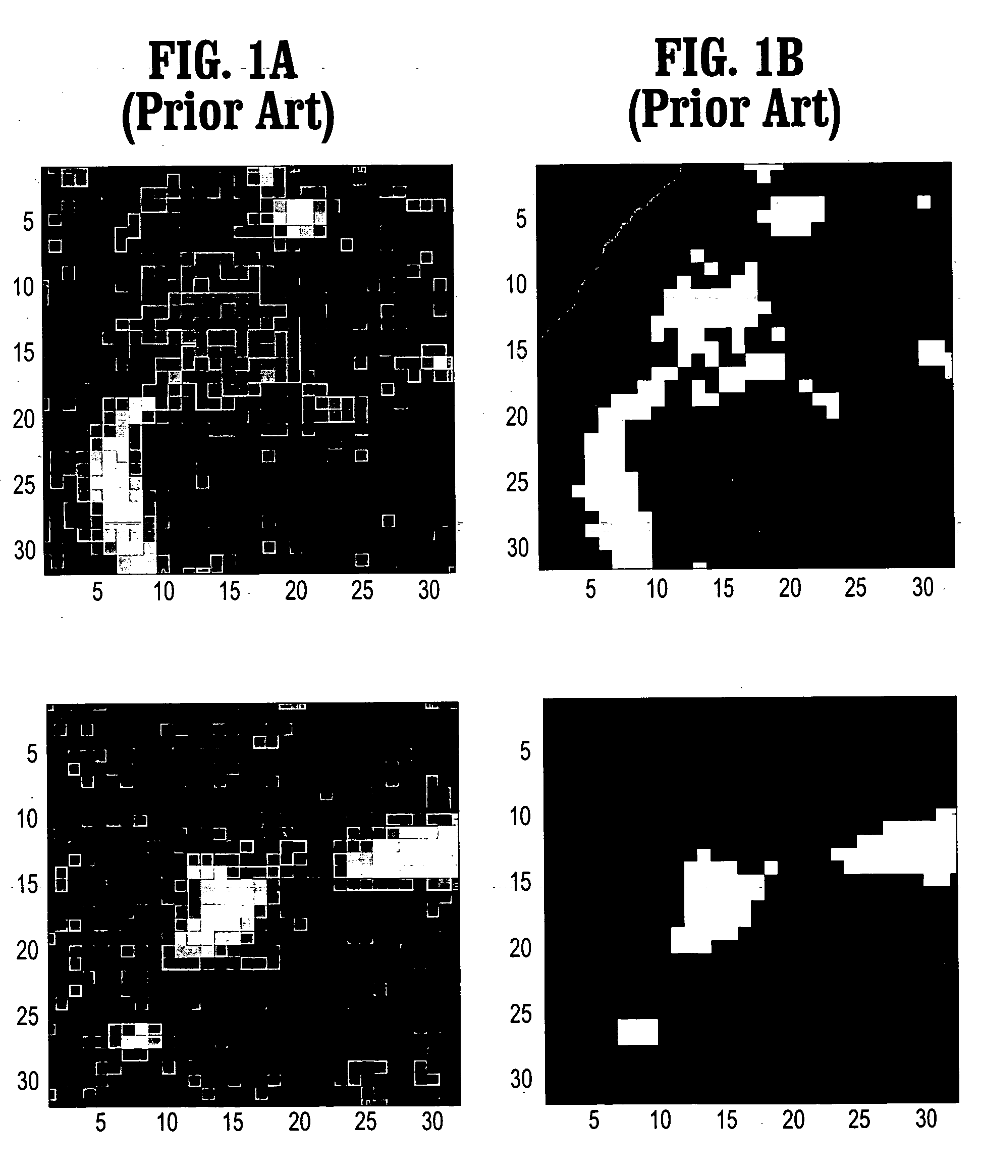
Although such solutions are sufficient to delineate the well-defined solid nodules with similar average intensity, they provide unreliable segmentation for the part- or non-solid nodules, as shown in FIGS.
The intensity thresholding method has been shown to fail for the non-solid case.

Method used


Image

  • 3D segmentation of targets in multislice image
  • 3D segmentation of targets in multislice image
  • 3D segmentation of targets in multislice image

Examples


Embodiment Construction

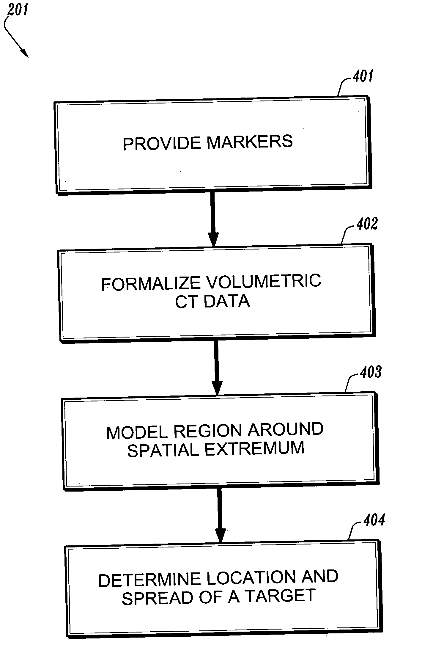
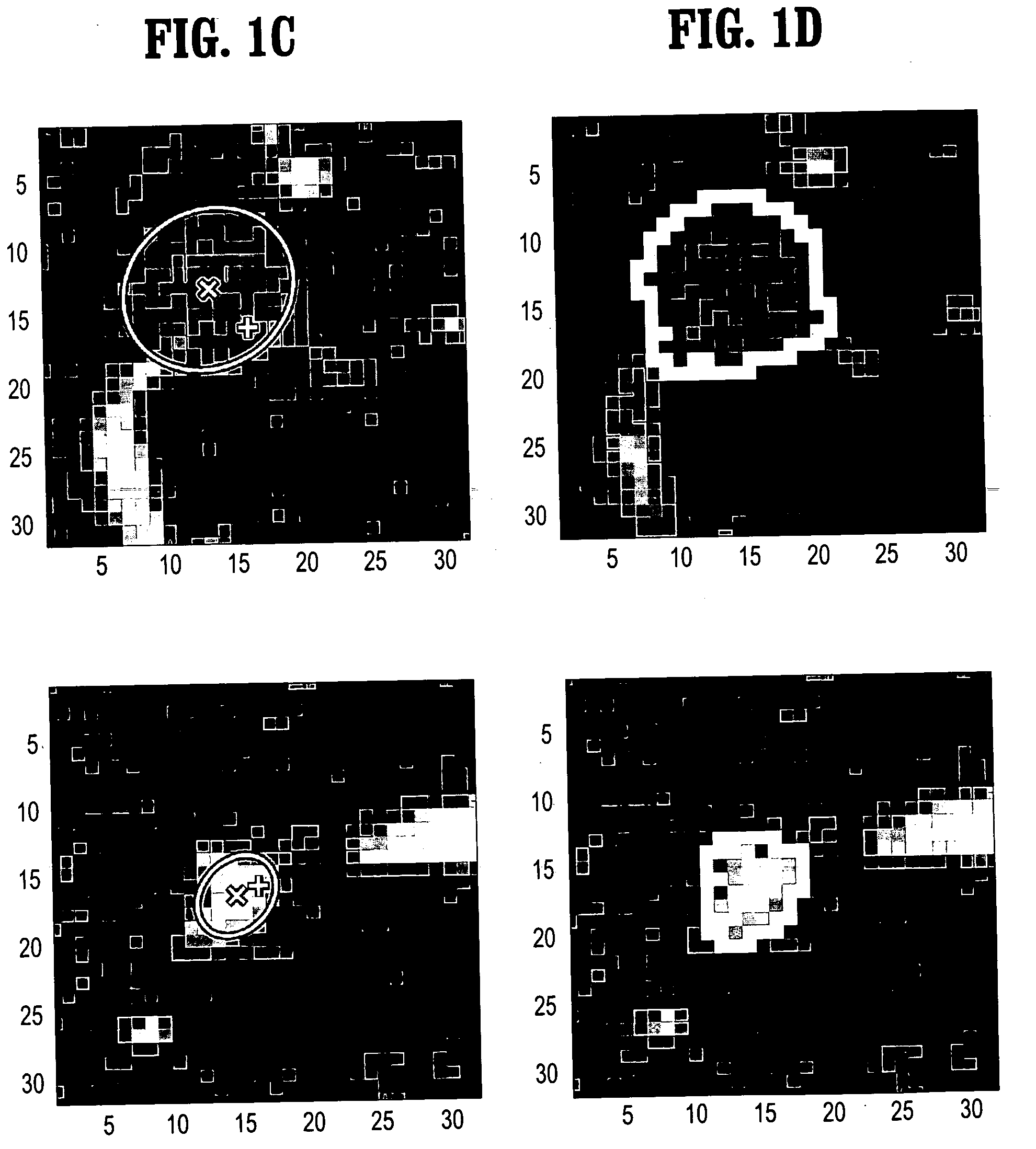
[0023] According to an embodiment of the present disclosure, a robust and accurate method for segmenting the 3D pulmonary nodules in multislice CT scans unifies the parametric Gaussian model fitting of the volumetric data evaluated in Gaussian scale-space and non parametric 3D segmentation based on normalized gradient (mean shift) ascent defining the basis of attraction of the target tumor in the 4D spatial-intensity joint space. This realizes the 3D segmentation according to both spatial and intensity proximities simultaneously. Experimental results show that the system and method reliably segment a variety of nodules including part- or non-solid nodules that poses difficulty for the existing solutions. The system and method also process a 32×32×32-voxel volume-of-interest efficiently by six seconds on average.

[0024] Referring to FIG. 2, the determination of 3D segmentation of volumes in multislice CT images includes 3D nodule center and spread estimation by fitting the anisotropi...


PUM


Abstract

A method for three-dimensional segmentation of a target in multislice images of volumetric data includes determining a center and a spread of the target by a parametric fitting of the volumetric data, and determining a three-dimensional volume by non-parametric segmentation of the volumetric data iteratively refining the center and spread of the target in the volumetric data.

Description

[0001] This application claims priority to U.S. Provisional Application Ser. No. 60 / 552,481, filed on Mar. 12, 2004, which is herein incorporated by reference in its entirety.BACKGROUND OF THE INVENTION [0002] 1. Field of the Invention [0003] The present invention relates to volumetric image data characterization, and more particularly to a system and method for 3D segmentation of targets in multislice images. [0004] 2. Description of Related Art [0005] One goal of the computer-assisted diagnosis with the chest CT scans (chest CAD) is reliable volumetric measurement of the pulmonary nodules. Tumor change qualification based on such volume measurements plays an integral part of the cancer therapy monitoring and post-surgical examinations. There are a number of previous studies addressing the computer-assisted volume measurement of nodules. In these studies, the 2D or 3D tumor segmentation based on voxel intensity thresholding is used as the foundation of their solutions. Although suc...

Claims


Application Information

Patent Timeline
IPC IPC(8): G06F19/00G06T5/00G06V10/26
CPCG06K2209/053G06K9/34G06V10/26G06V2201/032
Inventor OKADA, KAZUNORICOMANICIU, DORINKRISHNAN, ARUN
Owner SIEMENS MEDICAL SOLUTIONS USA INC
Learn More