Noninvasive method of ultrasound wound evaluation

a non-invasive method and ultrasound technology, applied in the field of ultrasound wound evaluation, can solve the problems of inconvenient measurement, contamination of the wound site, and the adhesive protective layer can damage the surrounding skin of the wound or further damage the wound itself, so as to improve the protection of the wound area, reduce the area of the wound, and improve the coverage of the wound area.

Inactive Publication Date: 2006-03-16
BIOSOUND
View PDF6 Cites 9 Cited by
  • Summary
  • Abstract
  • Description
  • Claims
  • Application Information

AI Technical Summary

Benefits of technology

[0008] The present invention overcomes the prior art by better protecting the wound by not utilizing an adhesive layer on the ultrasound media film. The present invention also allows for better coverage of the wound area by not restricting the size of the film layer by the adhesive edges. The invention uses more accurate measurement techniques to estimate the volume of a wound than previous methods.
[0009] The invention comprises a method for evaluating the extent of wounds, using a non-adhesive based couplant layer and ultrasound for image acquisition and applying a computer based measurement package of an ultrasound equipment system to measure either the wounds linear parameters, or the circumference and depth for volume determination. This information is used for the medical specialist to evaluate the efficacy of a particular treatment in the reduction of the wound area.

Problems solved by technology

This resulted in an imprecise measurement.
While these methods have proven to be effective, the adhesive protective layer can damage the surrounding skin to the wound or further damage the wound itself.
Other methods of applying ultrasonic waves to analyze wounds by not covering the wound can result in contamination of the wound site or ineffective application of the ultrasound waves due to not having an effective coupling medium in which to receive the ultrasonic waves.
The drawback to this system is the adhesive barrier, which is contraindicated in weak, friable skin adjacent to ulceration.
In addition, the barrier is often too small to adequately protect the entirety of the lower extremity wounds.
Also the method of multiplying the length, width and depth of a wound to obtain a volumetric measurement is often inaccurate.
In addition, there is no protocol for the exact location (within the body planes) these measurements are to be taken.

Method used


Image

  • Noninvasive method of ultrasound wound evaluation
  • Noninvasive method of ultrasound wound evaluation
  • Noninvasive method of ultrasound wound evaluation

Examples


Embodiment Construction

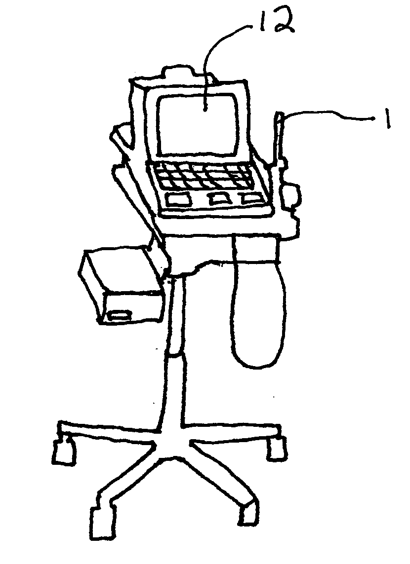
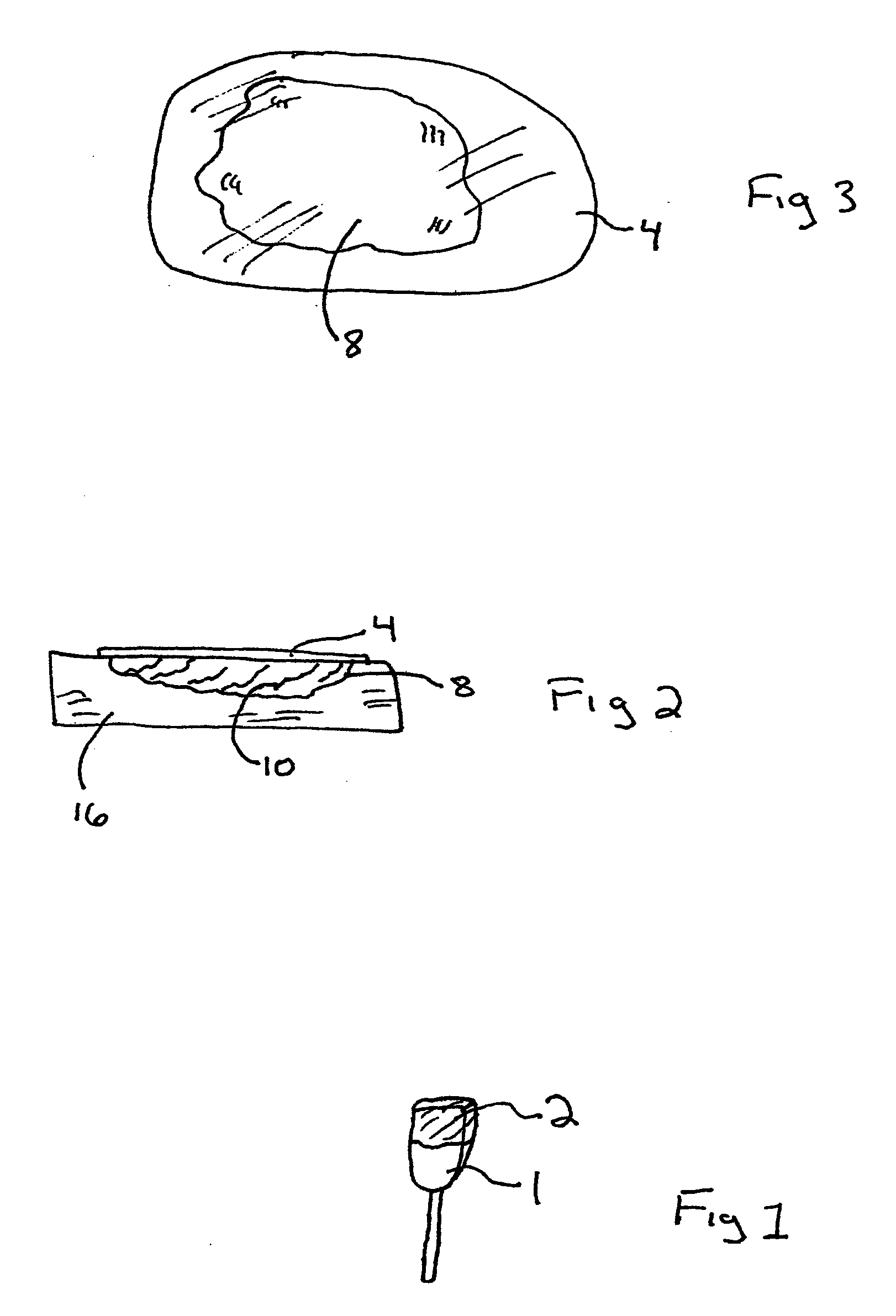
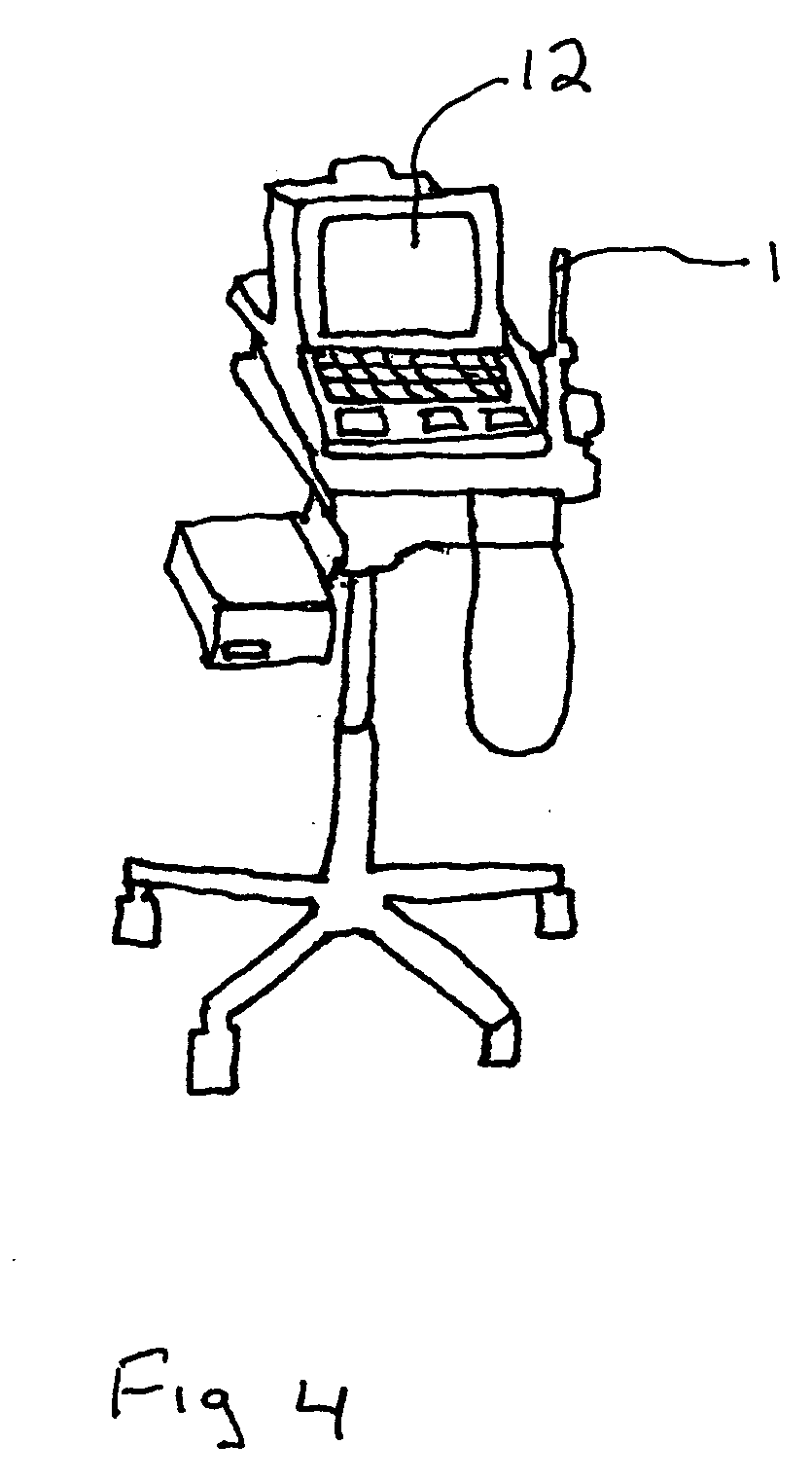
[0014] With reference to the drawings, and specifically FIGS. 1-4, The method uses a commercially available ultrasound couplant sheet 4, of sufficient size to completely cover a wound 8 in a human body after it is filled with a sterile coupling gel 10. A linear or convex array ultrasound transducer 1 is scanned over the covered wound 8 to image the extent of the wound 8. The cover 4 is not sealed with adhesive edges area, as there is a potential to cause damage to the sensitive skin and tissue layers 16 area adjacent to the wound. The cover 4 is held in place over the wound 4 by making contact with the coupling gel 10. To prevent contamination of the wound during the scan the ultrasound probe 1, which may be a broad bandwidth linear or convex array transducer, is covered with a commercially available sterile sheath 2. The ultrasound transducer 1, is connected to a computer controlled ultrasound system 12 which includes a keyboard, joystick, and other clinicain operated controls. The...


PUM


Abstract

A noninvasive method of ultrasound wound evaluation whereby a health care professional fills a wound with a sterile coupling gel, covers the wound with a non-adhesive couplant sheet, scans the wound with a ultrasound transducer that has been covered with a sterile sheath, thereby producing measurement data about the wound including the diameter, the perimeter and the depth, converting the data into a report package to be utilized by a health care professional to characterize the wound.

Description

PRIORITY [0001] The following non-provisional patent application claims priority to provisional application U.S. Ser. No. 60 / 602,732.FIELD OF THE INVENTION [0002] The present invention relates generally to the field of wound evaluation using ultrasound, and more specifically, to a method of measuring and evaluating a wound present on a human body by methodically following a procedure to protect and prepare the wound for analysis by a healthcare professional utilizing ultrasonic waves. BACKGROUND OF INVENTION [0003] Diagnostic ultrasound has proven to be an invaluable aid in the appreciation of the extent and damage caused by ulcerations. With deep, tunneling ulcerations, the method of clinically evaluating a wound was to insert a probe into the wound and “sound” the surfaces to get a general idea of the ulcers extent. This resulted in an imprecise measurement. With diagnostic ultrasound, the clinician can visualize the wound's extent on different body planes, measure the base of the...

Claims


Application Information

Patent Timeline
Patent Type & Authority Applications(United States)
IPC IPC(8): A61B8/12
CPCA61B5/1075A61B8/4281A61B8/0858
Inventor CHAPMAN, JAMES VINCENT
Owner BIOSOUND
Why Eureka
Try Eureka