
Visualization airway apparatus and methods for selective lung ventilation

a technology of airway apparatus and airway filter, which is applied in the field of airway filter apparatus and methods for selective lung ventilation, can solve the problems of double-lumen endobronchial tube and inability to maintain fiberoptic device within the endobronchial tube during the surgical procedur

Inactive Publication Date: 2007-09-20
EZC MEDICAL
View PDF41 Cites 79 Cited by
  • Summary
  • Abstract
  • Description
  • Claims
  • Application Information

AI Technical Summary

Benefits of technology

[0014] It is another object of the present invention to provide an airway device that can allow the operator to determine the placement of the airway device without the need to use a pediatric fiberscope or otherwise unduly obstruct the ventilation pathway.
[0015] It is a further object of the present invention to provide an airway device that allows the operator to monitor the position of the airway device as it is being used.
[0016] These and other advantages can be accomplished by providing an airway device having two lumens and a visualization device for allowing internal visualization of the placement of the airway and ongoing monitoring of the positioning of the device.
[0021] Observation of the imaging device may allow the user to determine whether the distalmost balloon becomes dislodged during the procedure through ongoing monitoring. These changes may indicate that the airway device is no longer properly positioned, thereby allowing the user to reposition the device before the patient suffers consequential harm. Additionally, the imaging device may be used during the initial positioning of the airway device to confirm proper placement as the airway device is inserted into the patient.

Problems solved by technology

Double-lumen endobronchial tubes are known in the art, but are also associated with known problems.
Due to the geometric limitations imposed by the size of the lumens and that of the fiberoptic device, it is not practical to maintain fiberoptic device within the endobronchial tube during the surgical procedure.

Method used


Image

  • Visualization airway apparatus and methods for selective lung ventilation
  • Visualization airway apparatus and methods for selective lung ventilation
  • Visualization airway apparatus and methods for selective lung ventilation

Examples


Embodiment Construction

[0032] The present invention is directed at a dual lumen airway device that comprises a visualization device that can assist in determining the placement of the device and identifying any subsequent repositioning. Accordingly, the user can ascertain the positioning of the device and continually monitor for inadvertent repositioning. The ability of the user to continually monitor the airway's position reduces the risk of an inadvertent repositioning remaining unnoticed.

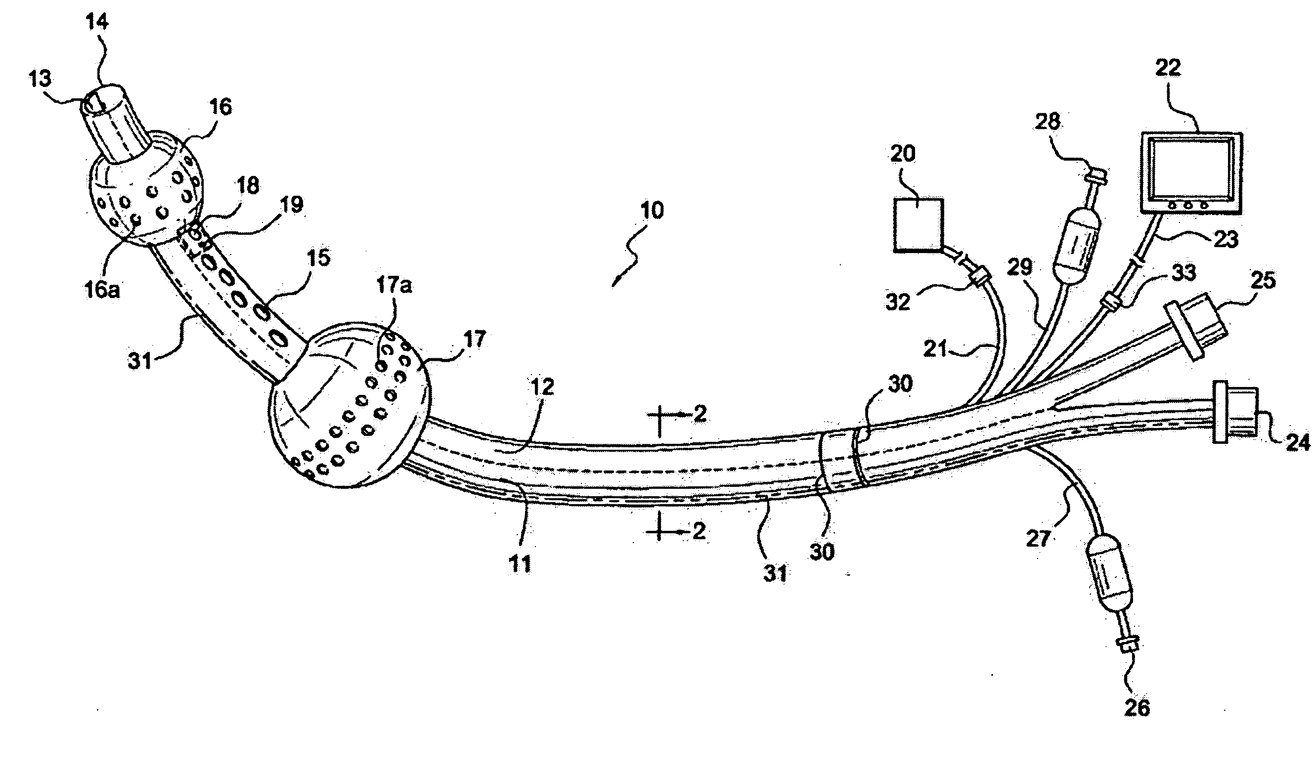
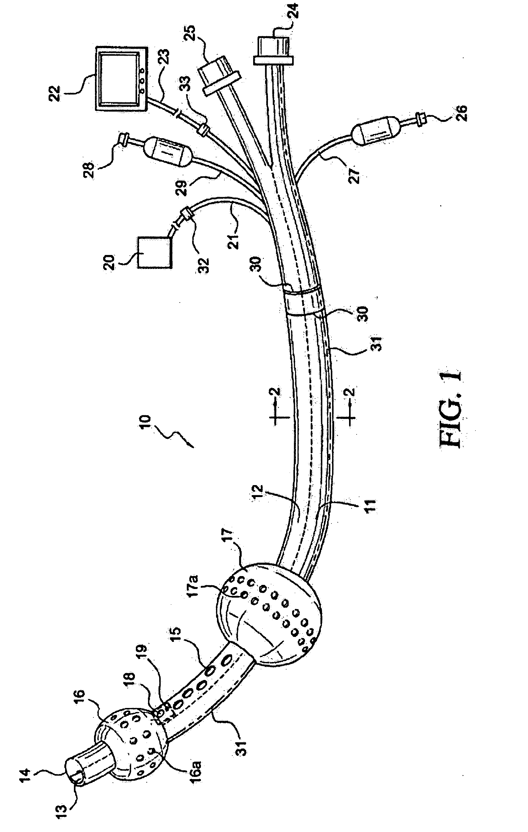
[0033]FIG. 1 depicts a preferred embodiment of the present invention that is appropriate for use in the upper airway and may be placed either in the patient's trachea or esophagus. Device 10 has tracheal lumen 11 and esophageal lumen 12. Aperture 13 of tracheal lumen 11 is located at distal end 14 of device 10. Apertures 15 of esophageal lumen 12 are located between distal balloon 16 and proximal balloon 17.

[0034] In this embodiment, balloons 16 and 17 comprise texture 16a and 17a. Texture 16a and 17a preferably comp...


PUM


Abstract

An airway device and method of use are provided, in which the airway device includes a dual lumen airway suitable for use as an endobronchial tube and having a sensor and a sensor monitoring device. In an embodiment of the device, the airway device includes imaging apparatus, optionally textured balloons, and image display device for monitoring the position of the device, thereby facilitating placement of the device and monitoring ability to determine a change in positioning.

Description

CROSS-REFERENCE TO RELATED APPLICATIONS [0001] This application is a continuation-in-part of patent application Ser. No. 11 / 303,343, filed Dec. 16, 2005.FIELD OF THE INVENTION [0002] The present invention relates to airway apparatus equipped with visualization capabilities and capable of providing selective ventilation to either of the lungs. BACKGROUND OF THE INVENTION [0003] In the medical profession, a patient may require surgery to treat a traumatic injury or a medical condition. If this surgery is to the heart, lungs, or other thoracic organs, a surgeon or other caregiver may have to access the organ by making an incision in the chest, known as a thoracotomy. [0004] When performing a thoracotomy, a patient is typically anesthetized and an airway device is inserted into the patient's trachea to allow for mechanical ventilation or other form of delivering oxygenating gasses to the patient's lungs. Under certain circumstances, such as for a lung surgery, the airway device is a dua...

Claims


Application Information

Patent Timeline
IPC IPC(8): A62B9/06A61M16/00
CPCA61B1/00082A61B1/00177A61B1/2676A61M16/04A61B1/0615A61M2205/32A61M16/0411A61M16/0409A61M16/0415A61M16/0436A61M16/0459A61M16/0486A61M16/0404A61M16/0477
Inventor GLASSENBERG, RAYMONDKIMMEL, ZEBADIAHSANDERS, GERALD J.
Owner EZC MEDICAL
Learn More