Scanned laser vein contrast enhancer

a contrast enhancer and laser vein technology, applied in the field of imaging devices, can solve the problem that no such control is possible, and achieve the effect of increasing the ability of the system, accurately detecting the position of blood vessels, and increasing the quality of the image captured

Active Publication Date: 2008-01-24
ACCUVEIN
View PDF28 Cites 54 Cited by
  • Summary
  • Abstract
  • Description
  • Claims
  • Application Information

AI Technical Summary

Problems solved by technology

In a camera based solution, since the infrared light floods the area being scanned, no such control is possible.

Method used


Image

  • Scanned laser vein contrast enhancer
  • Scanned laser vein contrast enhancer
  • Scanned laser vein contrast enhancer

Examples


Embodiment Construction

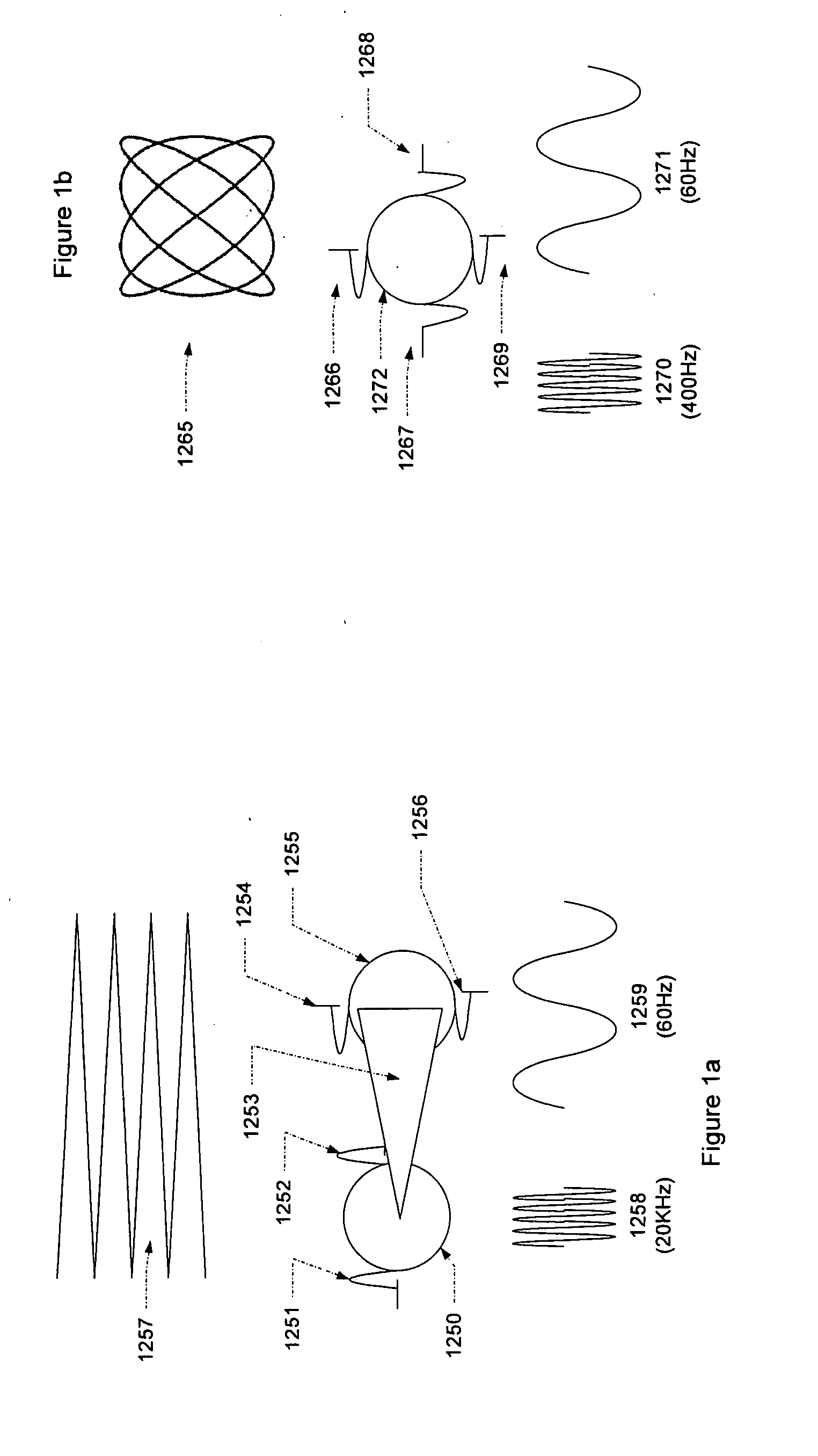
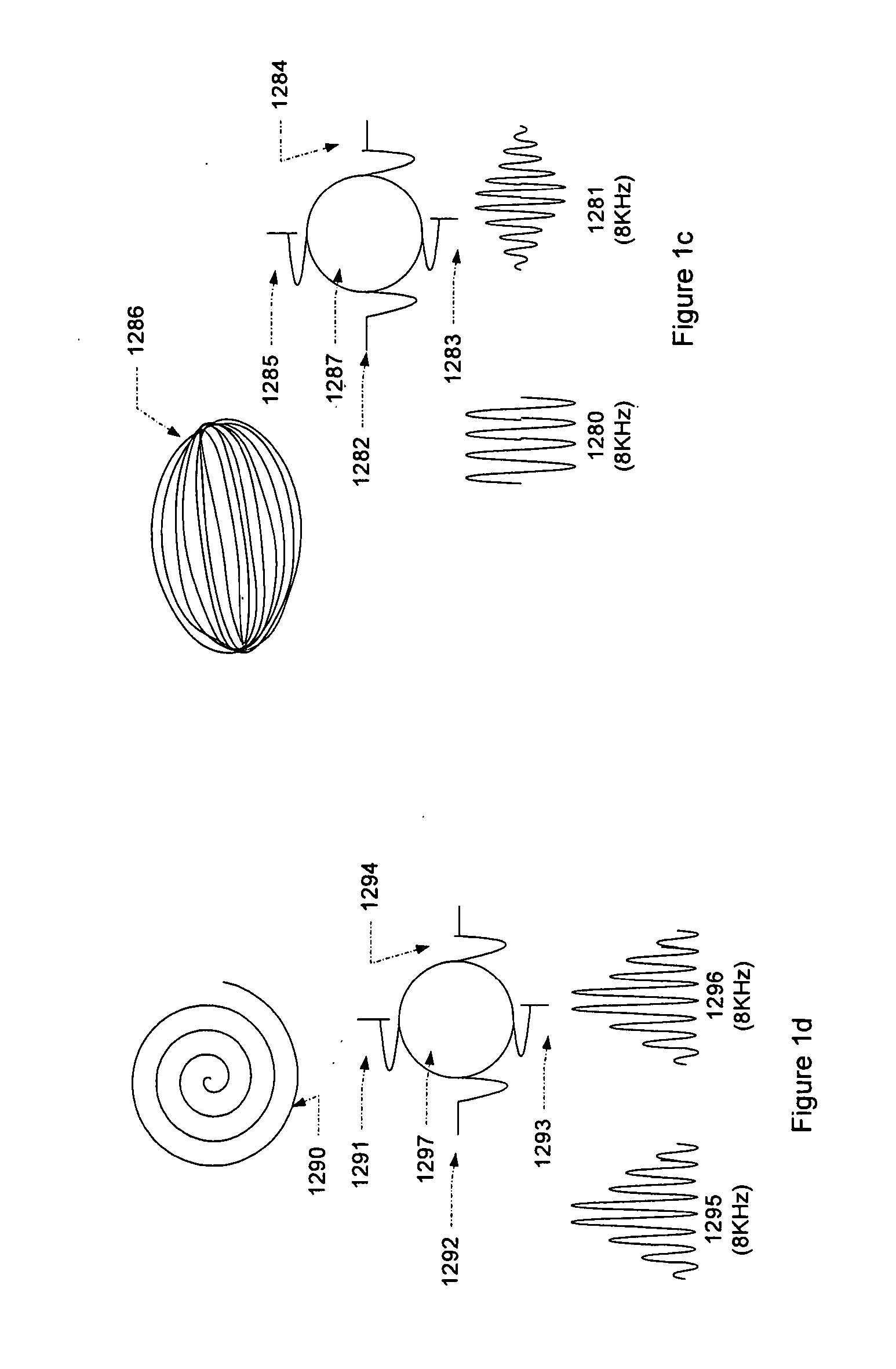
[0065] Referring to FIGS. 1a-1d, preferred embodiments are shown that generate raster, lissajous and collapsing ellipse and spiral patterns. The drawings show the mirror (e.g., 1250) on top of the electrical wave form (e.g., 1258) that is applied to the mirror to control its motion. These sinusoidal wave forms move the mirror in a back and forth pattern with maximum deflection at the peak of the wave, rest at the center of the wave and the opposite maximum deflection at the opposite peak of the wave. Also indicated in the drawing are the most preferred frequencies and the preferred ranges that would be used in an implementation of the invention.

Raster Pattern Generation

[0066] In FIG. 1a, a raster pattern 1257 is generated using two mirrors. Mirror 1250 is mounted so that it has a single degree of freedom of motion at fulcrum 1251 / 1252. A laser light source as described elsewhere strikes the mirror at an appropriate angle so that the angle of reflection is such that the light prop...


PUM


Abstract

The present invention is a Miniature Vein Enhancer that includes a Miniature Projection Head. The Miniature Projection Head may be operated in one of three modes, AFM, DBM, and RTM. The Miniature Projection Head of the present invention projects an image of the veins of a patient, which aids the practitioner in pinpointing a vein for an intravenous drip, blood test, and the like. The Miniature projection head may have a cavity for a power source or it may have a power source located in a body portion of the Miniature Vein Enhancer. The Miniature Vein Enhancer may be attached to one of several improved needle protectors, or the Miniature Vein Enhancer may be attached to a body similar to a flashlight for hand held use. The Miniature Vein Enhancer of the present invention may also be attached to a magnifying glass, a flat panel display, and the like.

Description

[0001] This application is a continuation in part of application Ser. No. 11 / 478,322, filed on Jun. 29, 2006 and also claims priority on provisional patent application entitled Three Dimensional Imaging of Veins, Application No. 60 / 817,623, also filed on Jun. 29, 2006, all disclosures of which are hereby incorporated by reference.FIELD OF INVENTION [0002] The invention described herein relates generally to an imaging device, in particular, an imaging means for enhancing visualization of components covered by living tissue thin opaque films. More particularly, the present invention is directed to enhancing the visualization of veins, arteries and other subcutaneous structures of the body for inter alia facilitating fluid insertion into or extraction from the body or otherwise visualizing subcutaneous structures for diagnosis of the medical condition of a patient or administration of medical treatment to a patient. BACKGROUND OF THE INVENTION [0003] A visit to a doctor's office, a cli...

Claims


Application Information

Patent Timeline
Patent Type & Authority Applications(United States)
IPC IPC(8): A61B6/00
CPCA61B5/489A61B5/0059A61B5/0062A61B2090/366
Inventor WOOD, FREDGOLDMAN, RONCONLON, STEPHENLUCIANO, VINCENT
Owner ACCUVEIN
Why Eureka
Try Eureka