Method and System For Regression-Based Object Detection in Medical Images

a regression-based object and image technology, applied in the field of object detection in medical images, can solve the problems of linear linear complexity of the classifier-based approach, extra computation of rotating images and computing their associated integral images, and difficulty in building a rapid detector for medical anatomy using the classifier-based approach

Active Publication Date: 2008-04-10
SIEMENS HEALTHCARE GMBH
View PDF8 Cites 22 Cited by
  • Summary
  • Abstract
  • Description
  • Claims
  • Application Information

AI Technical Summary

Problems solved by technology

In general, the computational complexity of the classifier-based approach linearly depends on the image size (for the translation parameter) and the number of tested orientations and scales.
Further, if the one classifier approach, which has been shown to perform better than the multiple-classifier approach, is used, then rotating the images and computing their associated integral images cost extra computations.
Therefore, due to the exhaustive nature of the classifier-based approach, it is challenging to build a rapid detector for medical anatomy using the classifier-based approach.

Method used


Image

  • Method and System For Regression-Based Object Detection in Medical Images
  • Method and System For Regression-Based Object Detection in Medical Images
  • Method and System For Regression-Based Object Detection in Medical Images

Examples


Embodiment Construction

[0023] The present invention is directed to a regression method for detecting anatomic structures in medical images. Embodiments of the present invention are described herein to give a visual understanding of the anatomic structure detection method. A digital image is often composed of digital representations of one or more objects (or shapes). The digital representation of an object is often described herein in terms of identifying and manipulating the objects. Such manipulations are virtual manipulations accomplished in the memory or other circuitry / hardware of a computer system. Accordingly, is to be understood that embodiments of the present invention may be performed within a computer system using data stored within the computer system.

[0024] An embodiment of the present invention in which a regression function is trained and used to detect a left ventricle in an echocardiogram is described herein. It is to be understood that the present invention is not limited to this embodi...


PUM


Abstract

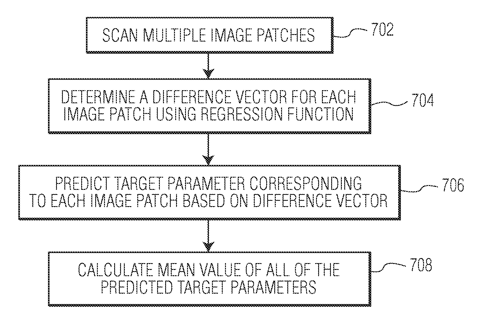
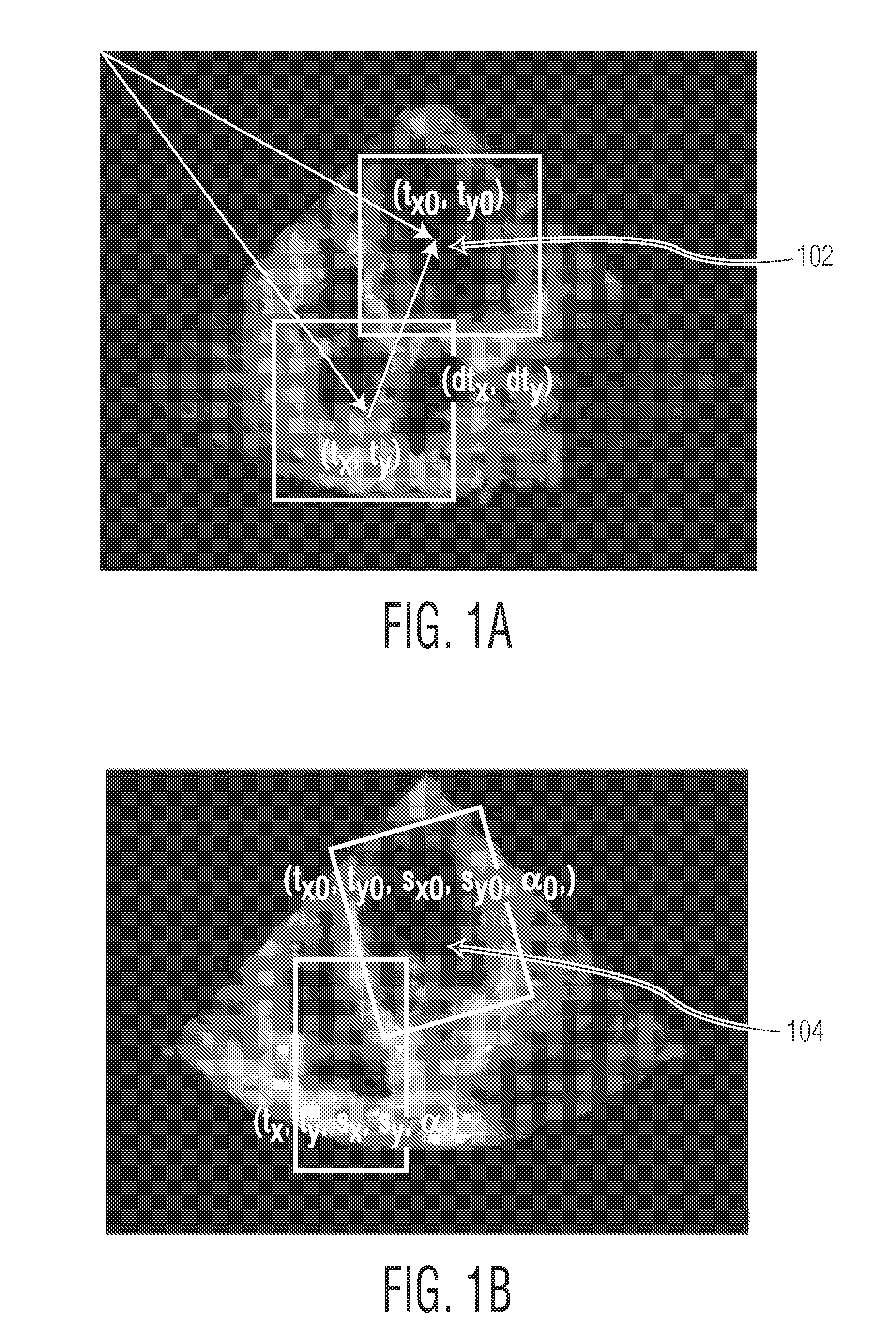
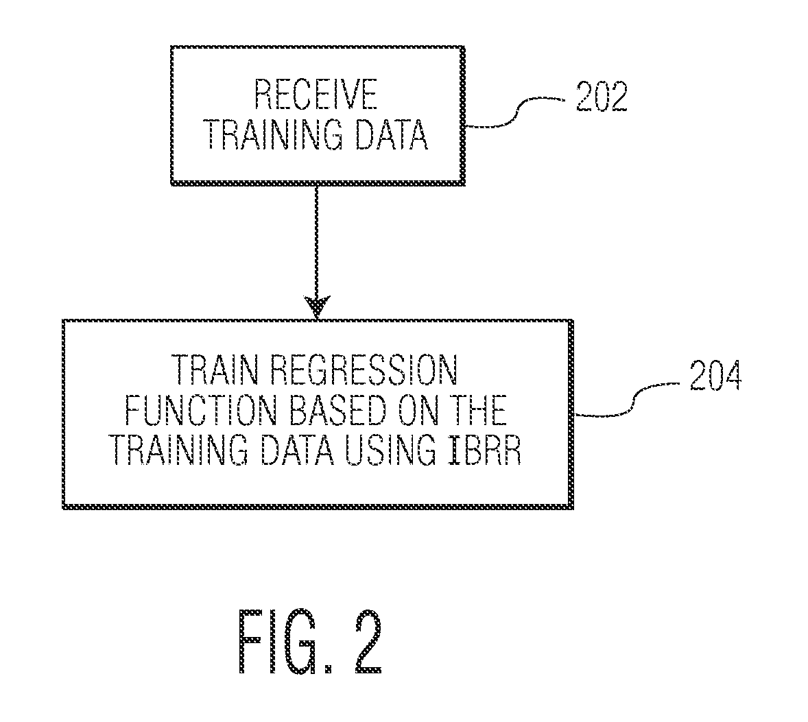
A method and system for regression-based object detection in medical images is disclosed. A regression function for predicting a location of an object in a medical image based on an image patch is trained using image-based boosting ridge regression (IBRR). The trained regression function is used to determine a difference vector based on an image patch of a medical image. The difference vector represents the difference between the location of the image patch and the location of a target object. The location of the target object in the medical image is predicted based on the difference vector determined by the regression function.

Description

[0001] This application claims the benefit of U.S. Provisional Application No. 60 / 849,936, filed Oct. 6, 2006, the disclosure of which is herein incorporated by reference.BACKGROUND OF THE INVENTION [0002] The present invention relates to object detection in medical images, and more particularly, to a regression method for detecting anatomic structures in medical images. [0003] Detecting anatomic structures in medical images, such as ultrasound, X-rays, CT images, MR images, etc., is important for medical image understanding. For example, in order to segment an anatomic structure in a medical image, conventional techniques typically involve anatomic structure detection followed by database-guided segmentation of the anatomic structure. Anatomic structure detection can also provide valuable initialization information for other segmentation techniques such as level set, active contour, etc. [0004] Conventional object detection techniques used to detect anatomic structures in medical i...

Claims


Application Information

Patent Timeline
Patent Type & Authority Applications(United States)
IPC IPC(8): G06K9/00G06V10/766G06V10/24G06V10/774
CPCG06K9/00G06K2209/05G06K9/72G06K9/6256G06V10/768G06V2201/03G06V10/24G06V10/766G06V10/7747G06V10/774G06F18/00G06F18/214
Inventor ZHOU, SHAOHUA KEVINZHOU, JINGHAOGEORGESCU, BOGDANCOMANICIU, DORIN
Owner SIEMENS HEALTHCARE GMBH
Why Eureka
Try Eureka