Devices, systems and methods for the collection, stimulation, stabilization, and analysis of a biological sample

a biological sample and system technology, applied in the field of biological sample collection, stimulation, stabilization, and devices, can solve the problems of unfixed/unstabilized samples, large number of facilities that routinely draw blood, and lack of equipment necessary to carry out conventional stimulation experiments

Inactive Publication Date: 2009-06-18
SMART TUBE INC
View PDF36 Cites 113 Cited by
  • Summary
  • Abstract
  • Description
  • Claims
  • Application Information

AI Technical Summary

Benefits of technology

[0006]Devices, systems, methods and kits for the collection, stimulation, stabilization and analysis of biological samples, including blood samples, are provided herein. To obtain the most physiologically and clinically relevant results from stimulation experiments performed on a biological sample, the biological sample should ideally be stimulated with a controlled dose of stimulus immediately after being obtained from a patient and, after a defined time interval of stimulation, the resulting intracellular signaling and / or gene transcription rapidly frozen in state by one or more stabilizing agents.

Problems solved by technology

A significant obstacle is that the majority of facilities that routinely draw blood lack the ability to carry out well-controlled stimulation experiments.
Specific problems include preparation of the stimulus, delivery of a precise amount of stimulus to the blood sample, and stabilizing the sample for later assessment of signaling state or transcript abundance.
Many of these facilities lack the equipment necessary to carry out conventional stimulation experiments.
Unfortunately, it is undesirable to ship certain samples in an unfixed / unstabilized state including blood samples positive for HIV or other infectious agents, and proper cryopreservation is also beyond the capabilities of many facilities.
Moreover, both cryopreservation and live shipping have been shown to induce changes in intracellular signaling and gene transcription and yield results that have been shown to poorly reflect the biology of blood cells in their native context.
These existing sample collection containers are not capable of executing multi-step experiments unless the user employs liquid handling devices that are not available at most locations where blood is drawn.

Method used


Image

  • Devices, systems and methods for the collection, stimulation, stabilization, and analysis of a biological sample
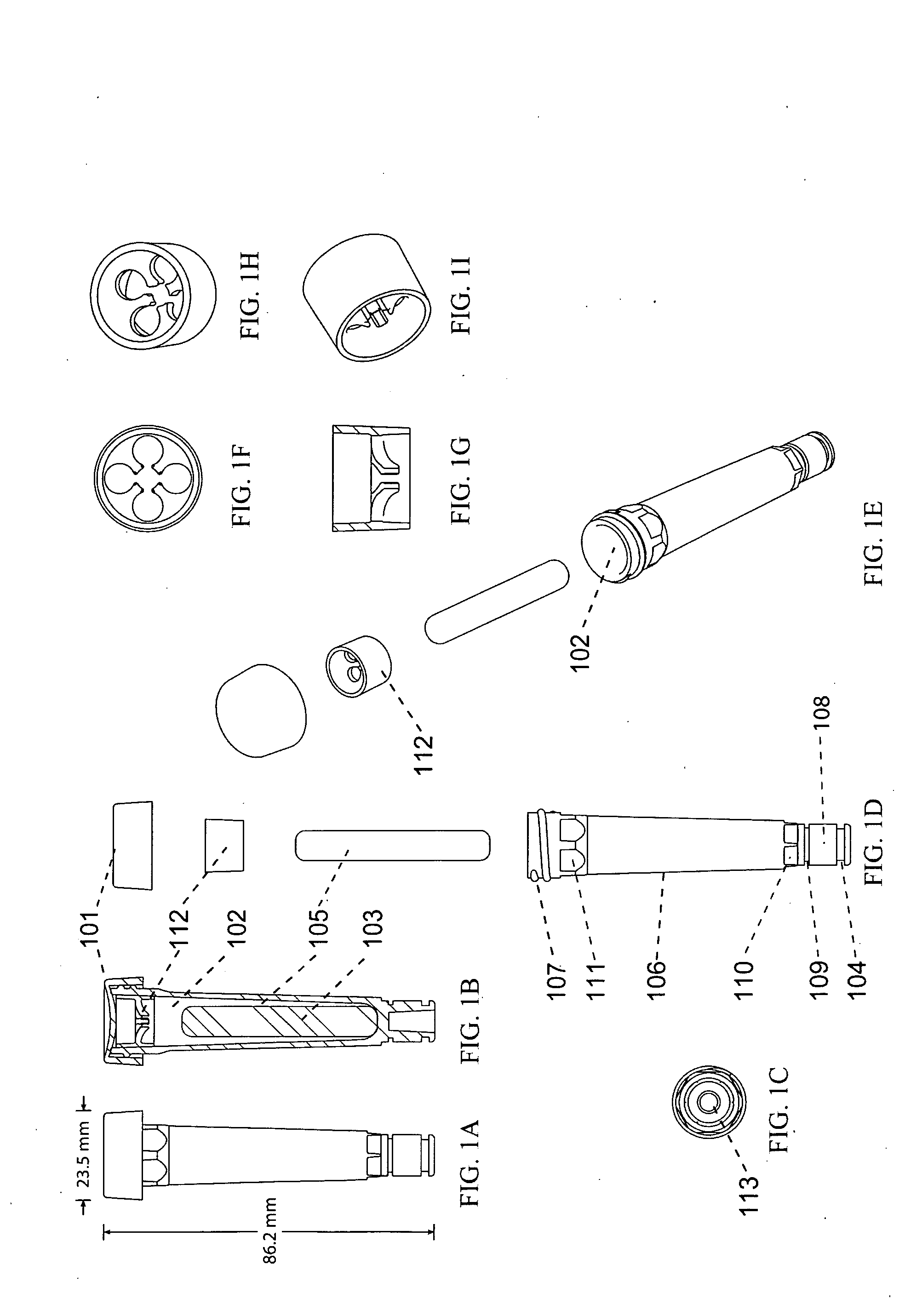
  • Devices, systems and methods for the collection, stimulation, stabilization, and analysis of a biological sample
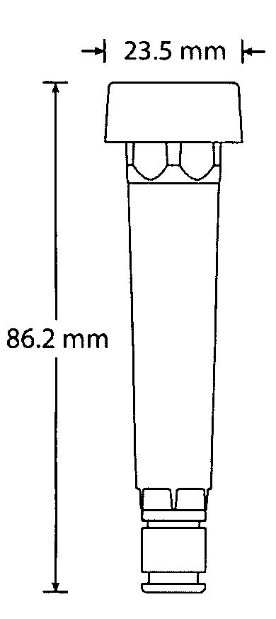
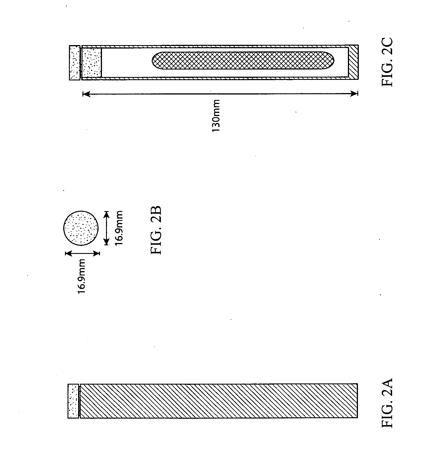
  • Devices, systems and methods for the collection, stimulation, stabilization, and analysis of a biological sample

Examples


example 1

Analysis of Stabilized Biological Sample Using Phospho-Specific Flow Cytometry

[0126]One process for analysis by phospho-specific flow cytometry includes the following steps. Frozen samples can be washed two times with ddH2O at physiological pH that may include an agent for lysing remaining erythrocytes if the biological sample was blood. Optionally, 0.1% Triton X100 or 0.1% saponin can be added to the ddH2O used to lyse the erythrocytes, detergents that have been shown to be effective for lysing erythrocytes. The cells are then washed with phosphate buffered saline and the pellet resuspended in 2 milliliters of a solution of 80% methanol and 20% phosphate buffered saline chilled to 4 degrees Celsius. The methanol fixed cell suspension can then be stored at −80 degrees Celsius. To continue processing the methanol fixed cell suspension is washed 2 times with staining media consisting of 0.5% bovine serum albumin dissolved in phosphate buffered saline and then stained and analyzed by p...

example 2

Analysis of Stabilized Biological Sample Using the Smart Tube Kit for Processing Samples Frozen in Smart Tubes, with Subsequent Analysis by Phospho-Specific Flow Cytometry

[0127]The following protocols uses the described container apparatus and Kit for processing samples frozen in Smart Tubes for subsequent analysis by phospho-specific flow cytometry.

[0128]Components of the Processing Kit include:[0129]i. Filter Cap (size of filter mesh openings between 500 microns and 2000 microns)[0130]ii. Lysis Buffer 1: 0.03% Tween 20 in double distilled H2O (ddH2O)[0131]iii. Lysis Buffer 2: 0.03% Tween 20 in 2× phosphate buffered saline (2×PBS)[0132]iv. One Liter of 2×PBS=16 g NaCl, 0.4 g KCl, 2.88 g Na2HPO4, 0.48 g of KH2PO4, and has a pH of 7.4.[0133]v. Permeabilization Buffer 1: 80% methanol with 20% PBS. (pre-chill on ice before use)[0134]vi. Staining Buffer 1: 0.5% bovine serum albumin in PBS

A. Thawing Collected, Stimulated, Stabilized Whole Blood Samples; Lysing Erythrocytes:

[0135]Thaw sam...


PUM

PropertyMeasurementUnit
angleaaaaaaaaaa
temperatureaaaaaaaaaa

Abstract

Devices, systems, methods and kits for the collection, stimulation, stabilization and analysis of biological samples, including blood samples, are disclosed. An embodiment of the invention includes a container having a side wall, a bottom wall and a closure member defining an internal compartment having arranged therein a partition defining and fluidly separating first and second chambers in the internal compartment, the first chamber positioned in association with the closure member to receive the biological sample; in which at least one wall is constructed of an elastically deformable material; in which the first chamber contains at least one stimulating agent; in which the second chamber contains at least one stabilizing agent; and in which the first and second chambers can be placed in fluid communication by a user without opening or otherwise compromising the fluid integrity of the internal compartment.

Description

CROSS-REFERENCE[0001]This application claims the benefit of U.S. Provisional Application No. 60 / 990,626, filed Nov. 28, 2007, which application is incorporated herein by reference in its entirety.BACKGROUND OF THE INVENTION[0002]Conventional diagnostics have focused on measuring the unperturbed biological state of a sample. In recent years there has been growing interest in exposing patient material to stimulatory agents such as cytokines, immunomodulatory factors, existing drugs, and new drug candidates, and then measuring the changes that have been induced in numerous cellular parameters such as intracellular signal transduction and genome-wide transcription. Several studies have shown that interrogating patient samples with stimuli reveals otherwise invisible biological states that have substantial clinical and diagnostic value (Irish, J. M. et al. Single cell profiling of potentiated phospho-protein networks in cancer cells. Cell (2004) 118, 217-28; Van Meter, M. E. et al. K-Ras...

Claims


Application Information

Patent Timeline
Patent Type & Authority Applications(United States)
IPC IPC(8): C12Q1/02C12M1/00C12M1/34G01N33/48G01N33/53C12Q1/68C40B30/04A01N1/02G01N27/26
CPCA61J1/2093A61J2001/202A61J2001/2027B01L3/505G01N2035/00782B01L2400/0481G01N1/38G01N2035/00465B01L2200/16A61J1/2027A61J1/065A61J1/05B65D25/08G01N33/5304
Inventor HALE, MATTHEW
Owner SMART TUBE INC
Why Eureka
Try Eureka