
Torc polynucleotides and polypeptides and method of use
a technology of polypeptides and polynucleotides, applied in the field of polynucleotides and polypeptides, cells, to achieve the effects of inhibiting development, inhibiting development, and inhibiting developmen
- Summary
- Abstract
- Description
- Claims
- Application Information
AI Technical Summary
Benefits of technology
Problems solved by technology
Method used
Image

Examples
example 1
Subcellular Localization of TORC1 Protein
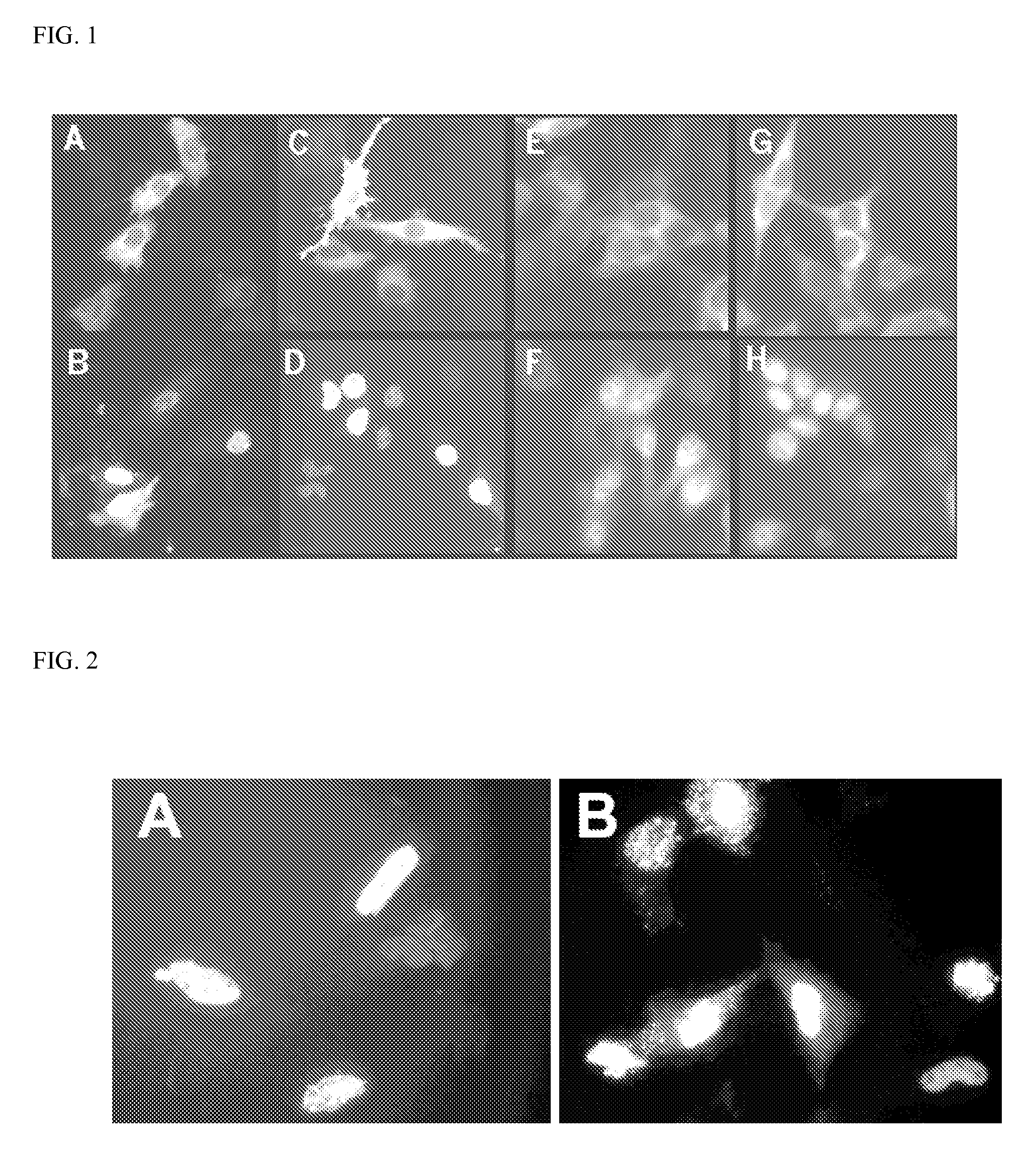
[0289]HeLa cells are transfected with FLAG-TORC1 (FIG. 1, panels A and B) or TORC1-eGFP (panels C and D). Cells are either untreated (panels A and C) or treated with 10 nM leptomycin-B (LMB), a fungal inhibitor of CRM1 mediated nuclear export of proteins, for 90 min (panels B and D). FLAG-tagged protein is visualized by immunofluorescence while eGFP-tagged proteins are directly visualized by fluorescence. Although TORC proteins have been shown to be CREB1-coactivators (Tourgenko et al., 2003), protein immunofluorescence using the FLAG epitope is found to be present predominantly in the HeLa cytoplasm (FIG. 1, panel A). Similar results are also obtained using direct fluorescence from eGFP with a TORC1eGFP fusion protein (TORC1-eGFP; FIG. 1, panel C). Since a variety of signal transduction proteins are regulated by either nuclear export or stimulus induced import, the localization of TORC1 is examined after treatment with LMB. After 90 minute e...
example 2
Subcellular Localization of TORC2 and TORC3 Proteins
[0290]The localization of human TORC2 and TORC3 is also examined. Both HeLa cells and HEK293 cells are transfected with either pCMV-TORC2-eGFP or pCMV-TORC3-eGFP. When expressed as eGFP fusions, TORC2 and TORC3 occurred constitutively in the nucleus in HeLa cells (FIG. 2). However, when expressed in HEK293 cells both TORC2- and TORC3-eGFP fusions proteins are largely, though not exclusively, present in the cytoplasm (FIG. 1, panels E and G, respectively, which show fluorescence primarily outside the nucleus). As with TORC1 in HeLa cells (Example 1), LMB treatment induces accumulation of both proteins in the nuclei of the cells, which now appear bright with fluorescence (FIG. 1, panels F and H, respectively). It may be concluded that the subcellular localization of the three human TORCs is regulated by nuclear export. (The localization of the TORC proteins in Examples 1 and 2 does not appear to be an artifact of either transfection ...
example 3
Identifying cDNA's that Induce TORC1 Nuclear Accumulation
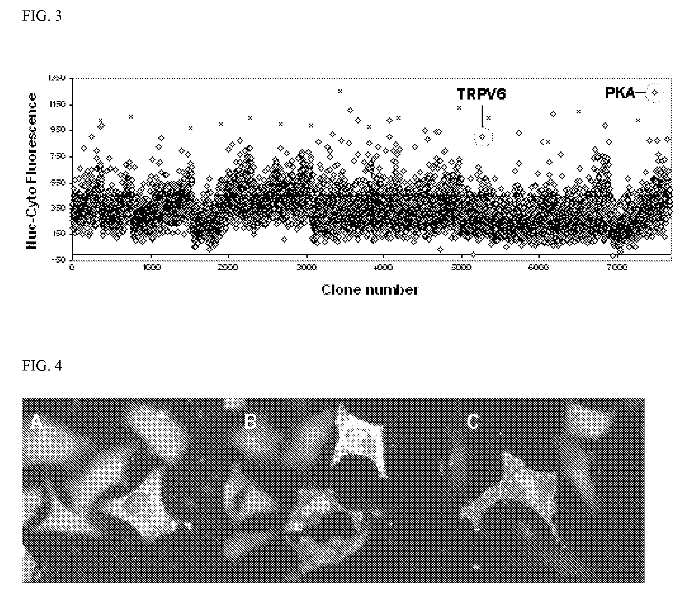
[0291]To identify cellular signals that regulate TORC translocation, a high-complexity screen is developed to identify genes that cause TORC1-eGFP to accumulate in the nucleus. HeLa cells are cotransfected with TORC1-eGFP in combination with approximately 7,000 individual cDNA expression constructs from the MGC Full-length collection (Strausberg et al., 2002). Forty-eight hours after transfection, the cells are fixed and the relative amounts of eGFP fluorescence in the cytoplasm and the nucleus are determined using the Cellomics ArrayScan II automated microscope. To provide a positive control for TORC1-eGFP translocation, one well in each 384-well cell-culture plate is treated with LMB prior to fixation. Translocation is monitored by plotting the nuclear-cytoplasmic fluorescence difference. Translocation via LMB is detected in virtually all control wells (FIG. 3, “X”). Potentially active cDNA's are recovered from clones with h...
PUM
| Property | Measurement | Unit |
|---|---|---|
| Fraction | aaaaa | aaaaa |
| Concentration | aaaaa | aaaaa |
| Biological properties | aaaaa | aaaaa |
Abstract
Description
Claims
Application Information