Dissecting cannula and methods of use thereof

a technology of dissection cannula and cannula, which is applied in the direction of surgical staples, catheters, osteosynthesis devices, etc., can solve the problems of reducing the number of devices that a physician must manipulate in the surgical area, reducing the amount of tissue dissection that a physician typically has less control over, and the device is still not optimal

Active Publication Date: 2009-12-17
ATRICURE
View PDF99 Cites 72 Cited by
  • Summary
  • Abstract
  • Description
  • Claims
  • Application Information

AI Technical Summary

Benefits of technology

[0015]Descriptions of the varying dissection modes are discussed below. However, the varying modes allow for gentle dissection, gradual dilation dissection, as well as a more forceful dissection by expansion. The latter dissection mode is useful to create a cavity between adjacent body structures or organs.

Problems solved by technology

Clearly, doing so reduces the number of devices that a physician must manipulate in the surgical area as well as the number of devices that are advanced through the body opening / port / incision.
However, because this dissection relies upon somewhat uncontrollable expansion of the balloon (as the internal balloon pressure increases), the physician typically has less control over the amount of tissue dissection as compared to using a non-expanding structure to physically dissect tissue.
While obturator type devices avoid the problems with somewhat unpredictable dissection via balloon expansion, such devices are still not optimal.
In any event, the balloon dissection or dissection via obturator dilation as described above do not provide the physician with the ability to tease or loosen adjoining tissue for a more controlled dissection of tissue.
Another drawback with conventional devices is their failure to accommodate removal of debris that is generated by the tissue dissection process.
Such debris interferes with visualization through the scope.
Without the ability to irrigate the scope, a physician will be forced to repeatedly remove the scope from the surgical site and body for cleaning.
Removal and cleaning of the scope increases the length and therefore the risk associated with the surgical procedure.
Moreover, apart from the debris, in the obturator-type devices described above, the closed transparent end of the device often causes a distorted view of the working area.
However, the devices and methods have applicability to any region in the body apart from the thoracic cavity.

Method used


Image

  • Dissecting cannula and methods of use thereof
  • Dissecting cannula and methods of use thereof
  • Dissecting cannula and methods of use thereof

Examples


Embodiment Construction

[0038]Methods and devices described herein provide for improved manipulation of organs and / or instruments within the body by creating working spaces within the body and adjacent to a target site. While the following disclosure discusses devices and methods for use in the thoracic cavity, such methods and devices can be applied to various body portions outside of the thoracic cavity. The methods and devices may allow for direct visualization along regions of anatomic structures not attainable with conventional approaches.

[0039]Furthermore, the methods and devices described herein may be used in conjunction with, or as an alternative to the conventional approaches described herein. For example, while some surgical approaches and procedures described herein rely on entry through the diaphragm of a patient to access a regions of the thoracic cavity, the surgical approaches and procedures can be combined with various other access methods.

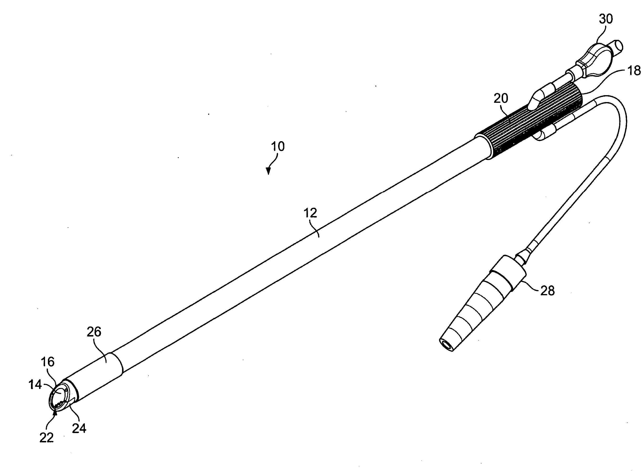
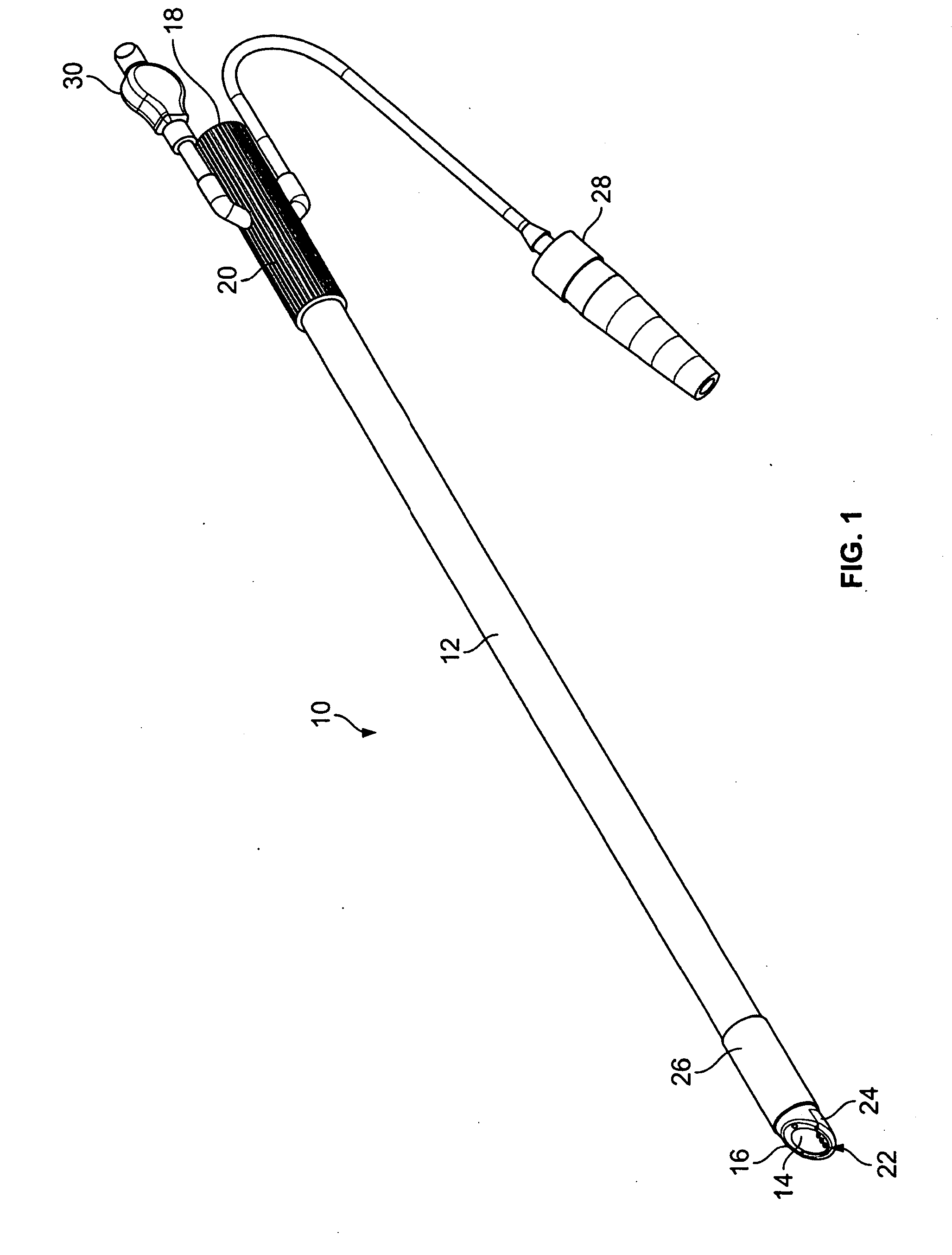
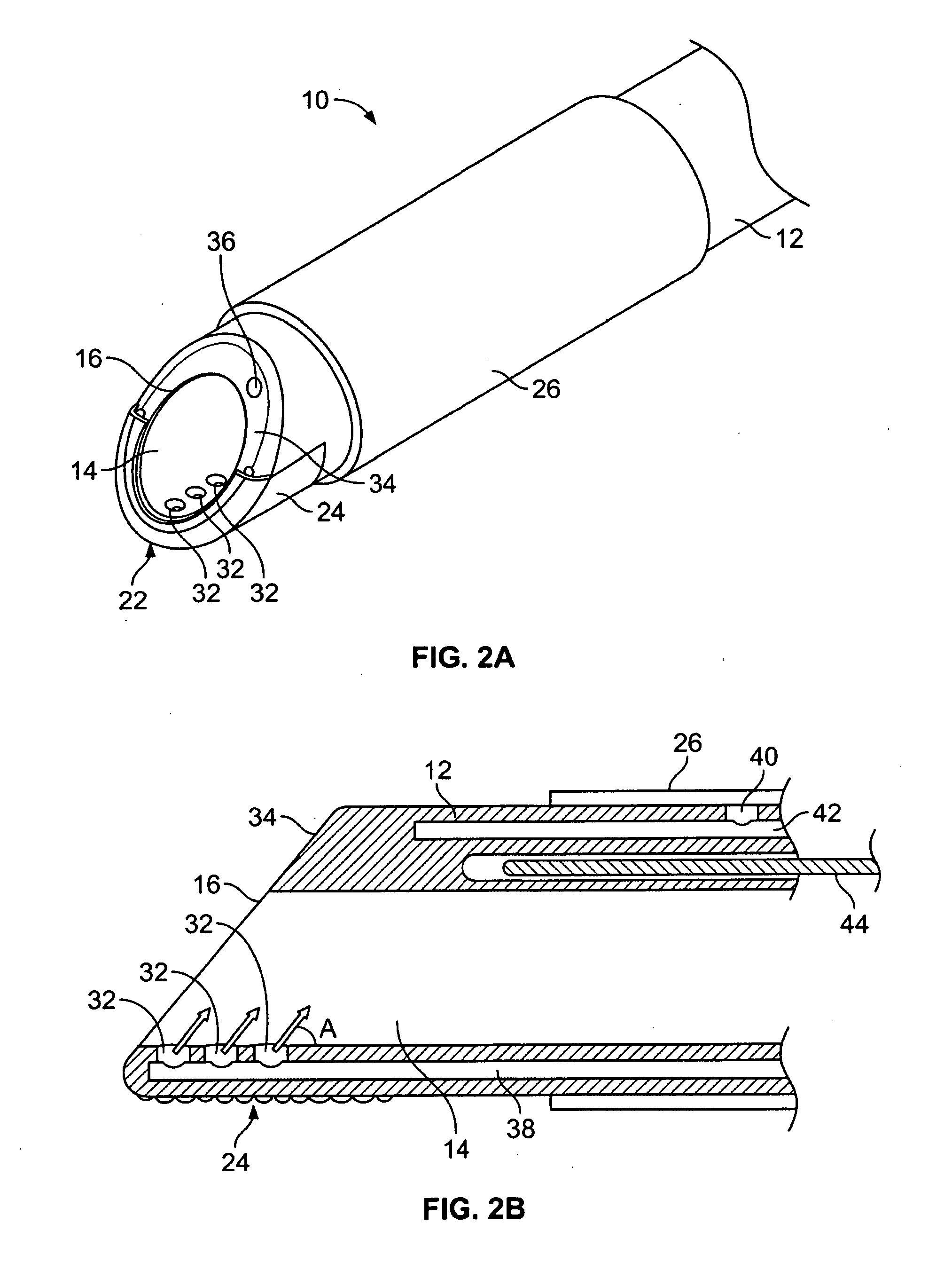
[0040]FIG. 1 shows one example of a tissue dissect...


PUM


Abstract

Methods and devices described herein facilitate improved access of locations within the body by providing a variety of dissection modes on a single access device.

Description

CROSS-REFERENCE TO RELATED APPLICATIONS[0001]The present application is a non-provisional of U.S. Provisional Patent Application No. 61 / 061,101, filed on Jun. 12, 2008, the content of which is incorporated herein by reference in its entirety.BACKGROUND OF THE INVENTION[0002]Methods and devices for access devices to allow improved manipulation of organs and / or instruments within the body by creating working spaces within the body and adjacent to a target site. The methods and devices can be used in various parts of the body. One particular application includes the use of the access devices and methods to advanced devices to a surface of the heart to create atrial lesion patterns one or more atrial surfaces of the heart. Scope based surgical tools (e.g., elongated cannula / tubular devices that allow viewing of internal body tissues) provide surgeons with an ability to view a surgical site through a lens / fiber optic / camera of the scope and also provide an ability to access the surgical ...

Claims


Application Information

Patent Timeline
Patent Type & Authority Applications(United States)
IPC IPC(8): A61B17/94
CPCA61B17/320016A61B2018/00589A61B17/3207A61B18/082A61B18/1492A61B19/24A61B19/5212A61B2017/00243A61B2017/22038A61B2017/22079A61B2017/320004A61B2017/320008A61B2017/320012A61B2017/320044A61B2017/320048A61B2017/3445A61B2018/00363A61B2018/00577A61B17/320068A61B90/02A61B90/361A61B2017/320069A61B2017/32007A61B2090/3614A61B2218/001
Inventor WHAYNE, JAMES G.FLEISCHMAN, SIDNEY D.SICVOL, CHRISTOPHER W.
Owner ATRICURE
Why Eureka
Try Eureka