
Intracardiac device for restoring the functional elasticity of the cardiac structures, holding tool for the intracardiac device, and method for implantation of the intracardiac device in the heart

a technology of intracardiac device and functional elasticity, which is applied in the field of intracardiac device for restoring functional elasticity of cardiac structures, can solve the problems of mechanical dysfunction, insufficient/uncompetence, hardening of attachment connections, etc., and achieve the effect of minimizing the reaction of the cardiac structure to the tissu

Active Publication Date: 2010-02-04
CUBE
View PDF27 Cites 150 Cited by
  • Summary
  • Abstract
  • Description
  • Claims
  • Application Information

AI Technical Summary

Benefits of technology

[0022]An object of the present invention is to minimize the tissutal reaction of the cardiac structure to a foreign body inside the heart.
[0023]Another object of the present invention is to reduce the thrombogenic capacity of the intracardiac device.
[0024]A further object of the present invention is to maintain the elastic property of the intracardiac device for a long period of time.

Problems solved by technology

The term heart failure defines a physiopathological state wherein the heart is not able to pump the blood in sufficient quantities for the metabolic requirements of the tissues, or wherein it performs a pumping action with excessively high filling pressure.
The mechanical function of each component of the cardiac pump (ventricular walls, structures that comprise the valves) has a physiologically elastic behaviour, and mechanical dysfunction is caused by the loss of elasticity of these components.
The most common form of heart failure of a mechanical nature is due to the excessive dilatation (dilative cardiomiopathy) of a ventricular chamber (in particular the left chamber) that can provoke a series of geometrical changes that also alter the geometry and function of the atrioventricular valve (above all the mitral valve) making it insufficient / not competent (valvulopathy).
Live experiments performed on sheep have shown a fibrotic reaction that in certain cases provoked attachment connection hardening, and consequential unwelcome stress on the sutures.
Furthermore, an excessive fibrotic reaction could reduce elasticity and compromise correct operating function.

Method used


Image

  • Intracardiac device for restoring the functional elasticity of the cardiac structures, holding tool for the intracardiac device, and method for implantation of the intracardiac device in the heart
  • Intracardiac device for restoring the functional elasticity of the cardiac structures, holding tool for the intracardiac device, and method for implantation of the intracardiac device in the heart
  • Intracardiac device for restoring the functional elasticity of the cardiac structures, holding tool for the intracardiac device, and method for implantation of the intracardiac device in the heart

Examples


Embodiment Construction

[0048]The Heart and the Cardiac Structures

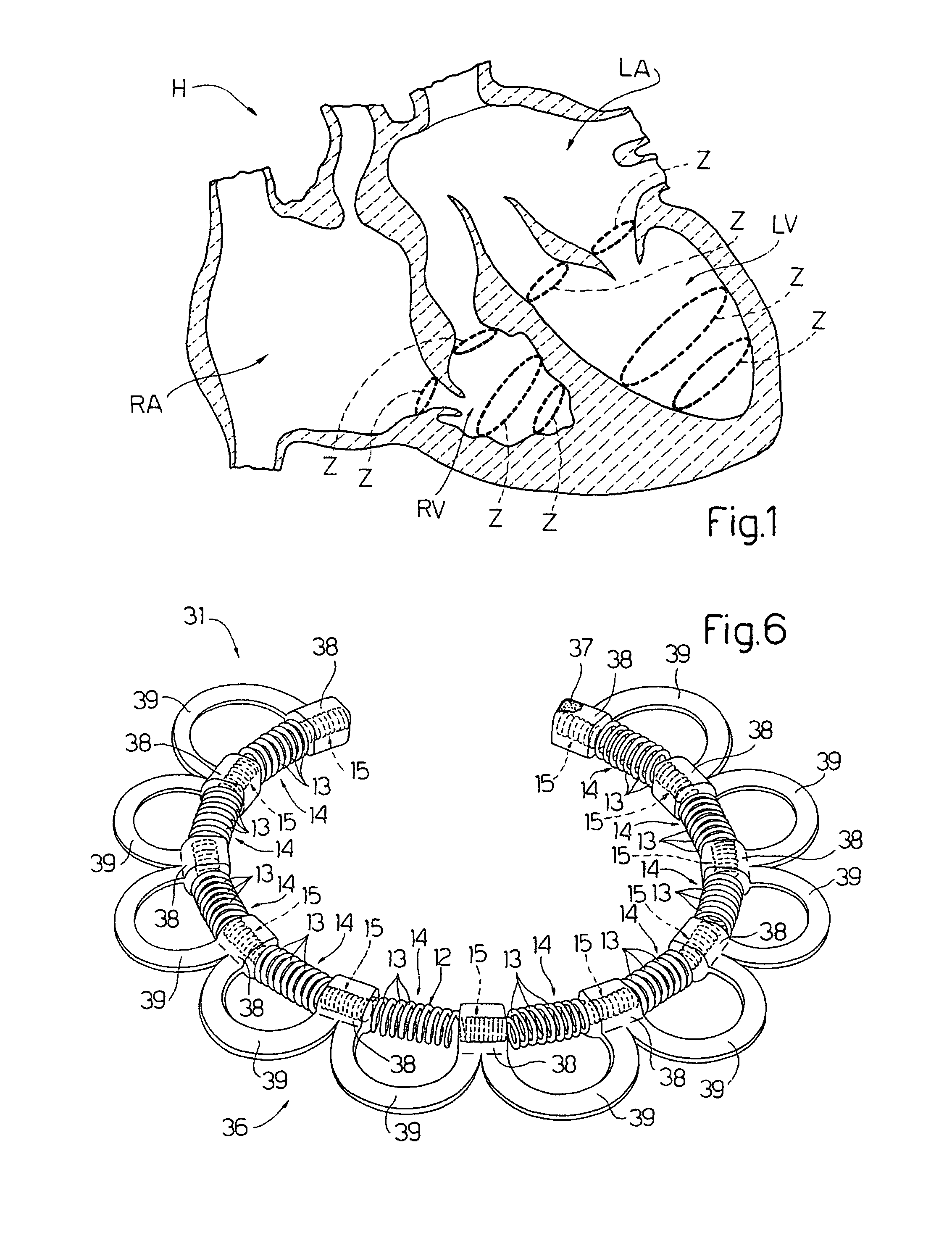
[0049]With reference to FIG. 1, H refers to a heart comprising four chambers: a left ventricle LV, a left atrium LA, a right ventricle RV, and a right atrium RA; four valves: mitral valve, aortic valve; tricuspid valve; and pulmonary valve.

[0050]Each ventricle is defined by a wall which expands and contracts to perform the pumping action, and each valve comprises an annulus.

[0051]In the present description, the term cardiac structure refers to both the ventricular walls and the valve annuli.

[0052]The dashed lines in FIG. 1 show the zones, Z, for possible connection of an intracardiac device object of the present invention, conceived for restoring cardiac structure function, should this result as not sufficient to completely perform the function for which it is conceived. Naturally, it is obvious that the zones Z shown herein are indicative and that the intracardiac devices can be implanted in zones other than those illustrated.

[0053]It is al...


PUM


Abstract

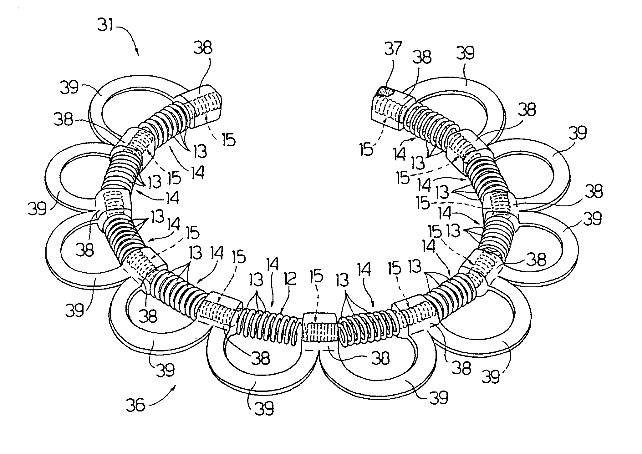
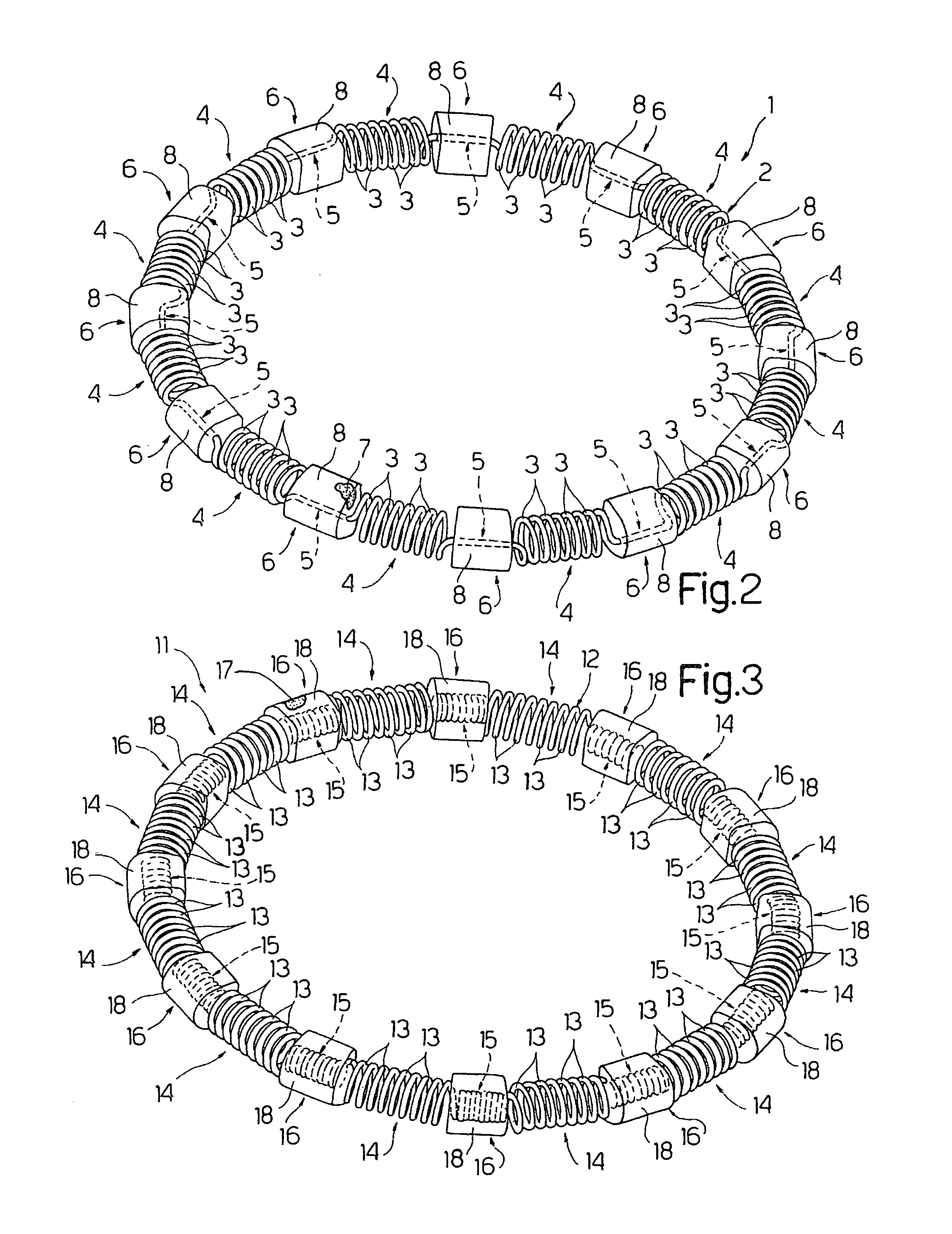
An intracardiac device for restoring the functional elasticity of the cardiac structures, in particular for the treatment of cardiomyopathies and or valvulopathies, by storing energy from the cardiac structures and ceding energy to the cardiac structures during the cardiac cycle, has an elongated shape, is at least partially wound in coils and is attachable to a cardiac structure; the coils are selected in material, number and dimension so as to allow an elastic elongation of the intracardiac device higher than 10% of the rest length of the intracardiac device and are exposed, in use, to the blood flow.

Description

FIELD OF THE INVENTION[0001]The present invention relates to an intracardiac device for restoring elasticity of cardiac structures.[0002]In particular, the present invention relates to a device for the treatment of cardiomyopathies and valvulopathies.BACKGROUND OF THE INVENTION[0003]In order to better understand cardiac dysfunctions such as cardiomyopathies and valvulopathies, object of the treatment and which affect the heart, we will provide a brief description of the heart structure and function.[0004]The heart comprises cavities which communicate with one another and with the venous and arterial system through valves. In the case in question, the heart comprises four cavities: a right atrium, a left atrium, a right ventricle, and a left ventricle; and four valves: the mitral valve, the aortic valve, the pulmonary valve and the tricuspid valve. The blood from the human body transits through the right atrium and from here crosses through the tricuspid valve, to the right ventricle...

Claims


Application Information

Patent Timeline
Patent Type & Authority Applications(United States)
IPC IPC(8): A61F2/00A61F2/04
CPCA61F2/2445A61F2/2466A61F2/2448A61F2/2481
Inventor FERRAZZI, PAOLO
Owner CUBE
Learn More