Method for detection of linear structures and microcalcifications in mammographic images

a linear structure and mammography technology, applied in image enhancement, image analysis, instruments, etc., can solve the problems of high rate of unnecessary biops, high rate of mcc fps, and inability to detect linear structures and microcalcifications in mammography images, so as to reduce mcc fps

Inactive Publication Date: 2010-04-29
CARESTREAM HEALTH INC
View PDF9 Cites 18 Cited by
  • Summary
  • Abstract
  • Description
  • Claims
  • Application Information

AI Technical Summary

Benefits of technology

[0012]The present invention is designed to overcome the problems set forth above. More particularly, with the present invention, all MCC candidates are assumed to be associated with linear structures until verified otherwise. Therefore, the present invention provides a method for detection of linear structures in mammography CAD systems with the objective of reducing MCC FPs.

Problems solved by technology

However, analyzing mammograms requires skilled radiologists whose performance can be degraded by the demand of viewing a large number of images in a finite amount of time.
It is very difficult to distinguish between malignant and benign microcalcification clusters (MCCs), even for experienced radiologists, which may lead to a high rate of unnecessary biopsies.
Their results suggest significant differences between the different approaches.
Moreover, Wai et al say that it is computationally inefficient to generate actual linear structures just for the purpose of confirming the association of an MCC candidate with a linear structure in mammography CAD.

Method used


Image

  • Method for detection of linear structures and microcalcifications in mammographic images
  • Method for detection of linear structures and microcalcifications in mammographic images
  • Method for detection of linear structures and microcalcifications in mammographic images

Examples


Embodiment Construction

[0031]The following is a detailed description of the preferred embodiments that use the current invention, reference being made to the drawings in which the same reference numerals identify the same elements of structure in each of the several figures.

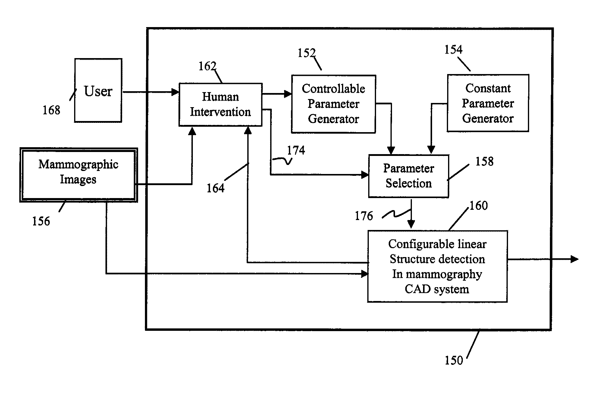
[0032]Turning now to FIG. 1, the method of the present invention will be outlined. FIG. 1 is a work flow chart 150 illustrating an embodiment that employs the linear structure detection method of the present invention. There are many variations in appearance of linear structures in mammograms in terms of contrast, brightness, texture and morphological shapes, among others. Therefore, in a practical image processing system it is desirable, as one measure, to synergistically integrate the skills of the human operator of the system with the power of the computer in the process of linear structure detection. A typical human excels in creativity, use of heuristics, flexibility and common sense; while a computer excels in speed of computatio...


PUM


Abstract

A method for linear structure detection in mammographic images, comprising: locating a plurality of microcalcification candidate clusters in digital mammographic images; extracting a region of interest that encloses each of the candidate clusters; processing the region of interest to generate feature points that reveal geometric properties in the region; applying a line detection algorithm to the feature points to produce a line model; and analyzing the line model to determine whether a true linear structure is present in the first region of interest.

Description

CROSS REFERENCE TO RELATED APPLICATIONS[0001]This application is a continuation-in-part of commonly assigned, copending U.S. patent application Ser. No. 11 / 834,243 filed Aug. 6, 2007 by Chen et al for LINE STRUCTURE DETECTION AND ANALYSIS FOR MAMMOGRAPHY CAD; and Ser. No. 12 / 058,803 filed Mar. 31, 2008 by Chen et al for LINEAR STRUCTURE VERIFICATION 1N MEDICAL APPLICATIONS. The disclosures of these applications hereby are incorporated by reference into the present specification.FIELD OF THE INVENTION[0002]The invention relates generally to image processing in medical applications, and in particular, to computer aided detection (CAD) in mammography.BACKGROUND OF THE INVENTION[0003]Mammography is an effective method of screening for breast cancer, a leading cause of mortality among women. However, analyzing mammograms requires skilled radiologists whose performance can be degraded by the demand of viewing a large number of images in a finite amount of time. A CAD feature has been prov...

Claims


Application Information

Patent Timeline
Patent Type & Authority Applications(United States)
IPC IPC(8): G06K9/00
CPCG06T7/0012G06T2207/30068G06T2207/20104G06T2207/10116
Inventor CHEN, SHOUPURAY, LAWRENCE A.
Owner CARESTREAM HEALTH INC
Why Eureka
Try Eureka