Genes and polypeptides relating to hepatocellular or colorectal carcinoma

a technology of hepatocellular or colorectal carcinoma and gene, applied in the field of biological science, can solve the problems of toxic to cancer cells and normal growing cells, and achieve the effect of inhibiting the expression of reporter genes

Inactive Publication Date: 2010-11-18
ONCOTHERAPY SCI INC
View PDF10 Cites 3 Cited by
  • Summary
  • Abstract
  • Description
  • Claims
  • Application Information

AI Technical Summary

Problems solved by technology

Many anticancer drugs, such as inhibitors of DNA and / or RNA synthesis, metabolic suppressors, and DNA intercalators, are not only toxic to cancer cells but also for normally growing cells.

Method used


Image

  • Genes and polypeptides relating to hepatocellular or colorectal carcinoma
  • Genes and polypeptides relating to hepatocellular or colorectal carcinoma
  • Genes and polypeptides relating to hepatocellular or colorectal carcinoma

Examples


example 1

Identification of Two Novel Genes, WDRPUH and KRZFDUH, Frequently Up-Regulated in HCCs

[0255]The expression profile of 20 HCCs were compared with that of corresponding non-cancerous liver tissues using in-house genome-wide cDNA microarray containing 23040 genes. More specifically, HCC tissues, and corresponding non-cancerous tissues were obtained with informed consent from surgical specimens of patients who underwent surgery. Total RNA was extracted from microdissected tissue with Qiagen RNeasy kit (Qiagen) or Trizol reagent (Life Technologies) according to the manufacturers' protocol. The extracted total RNA was treated with DNase I, amplified with Ampliscribe T7 Transcription Kit (Epicentre Technologies) and labeled during reverse transcription using Cy-dye (Amersham). Cy5 and Cy3 were used for labeling RNAs from non-cancerous tissue and tumor, respectively. Then, hybridization, washing, and detection were carried out according to the method of Ono et al. (Cancer Res 60: 5007-11 (...

example 2

Expression, Isolation, and Characterization of the Novel Human Gene WDRPUH

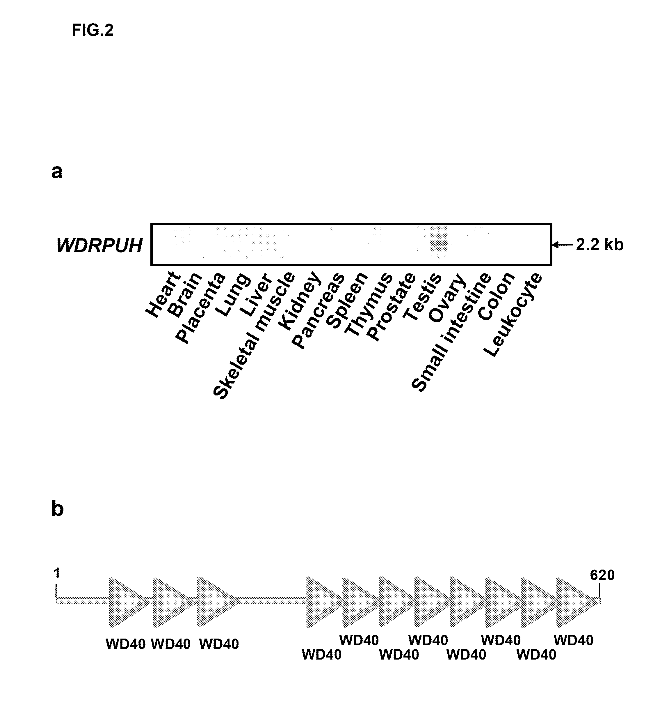
[0258]Next, multi-tissue northern blot analysis was performed using the PCR product of WDRPUH as a probe. More specifically, human multiple-tissue blots (Clontech) were hybridized with 32P-labeled PCR product of WDRPUH. Pre-hybridization, hybridization, and washing were performed according to the supplier's recommendations. The blots were autoradiographed with intensifying screens as −80° C. for 24 to 72 h. As a result, a 2-kb transcript was detected to be abundantly expressed in testis (FIG. 2a). Since D3197 was smaller than the WDRPUH cDNA detected on the Northern blot, next the inventors investigated the 5′ sequence of WDRPUH cDNA. First, the genomic sequence corresponding to D3197 was searched in genomic databases (http: / / www.ncbi.nlm.nih.gov / BLAST / ) using BLAST program to find a cosmid sequence (GenBank Accession No. AC026855) assigned to chromosomal band 17p13. Candidate-exon sequences of the genomic seq...

example 3

Subcellular Localization of WDRPUH

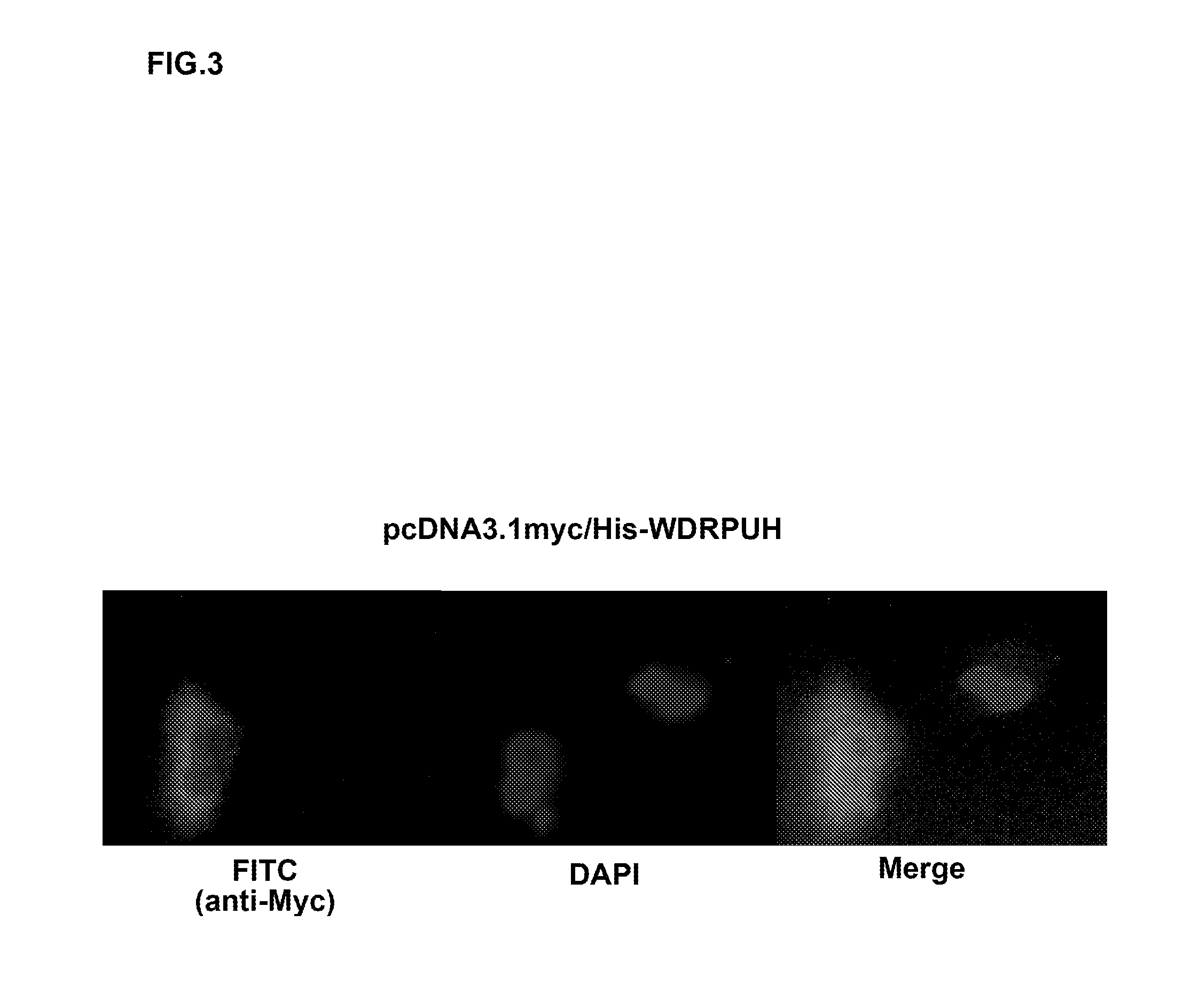
[0259]The entire coding region corresponding to WDRPUH was amplified using gene specific primer set: 5′-GGGGTACCACCATGGATAACAAAATTTCGCCGGAG-3′ [SEQ ID NO: 14] and 5′-CGGAATTCTCAGGAGGTATATGGGTACTTCCATGC-3′ [SEQ ID NO: 15]; and cloned into pcDNA3.1myc / His vector (Invitrogen). Then, the constructed vector pcDNA3.1myc / His-WDRPUH was transiently transfected into SNU475 cells (Korea cell-line bank) and the cells were grown in RPMI1640. The cells were fixed with PBS containing 4% paraformaldehyde for 15 min, then permeabilized with PBS containing 0.1% Triton X-100 for 2.5 min at room temperature (RT). Subsequently, cells were covered with 2% BSA in PBS for 24 h at 4° C. to block non-specific hybridization. 1:1000 diluted mouse anti-myc monoclonal antibody (Sigma) was used as the primary antibody for immunocytochemical staining, and the reaction was visualized after incubation with Rhodamine-conjugated anti-mouse secondary antibody (Leinco and ICN). Nuclei ...


PUM


Abstract

The present application provides novel human genes WDRPUH and KRZFPUH, and PPIL1 whose expression is markedly elevated in a great majority of HCCs and colorectal cancers, respectively, compared to corresponding non-cancerous tissues, as well as novel human gene APCDD1 whose expression is elevated in primary colon cancers and down-regulated in response to the transduction of wild-type APC1 into colon-cancer cells. The genes and polypeptides encoded by the genes can be used, for example, in the diagnosis of a cell proliferative disease, and as target molecules for developing drugs against the disease.

Description

[0001]The present application is a division of U.S. patent application Ser. No. 11 / 948,790, filed Nov. 30, 2007, which is a continuation of U.S. patent application Ser. No. 10 / 517,151, filed Aug. 18, 2005, which is a U.S. National Phase of International Application No. PCT / JP2003 / 07070, filed Jun. 4, 2003, which claims the benefit of U.S. Provisional Application No. 60 / 386,985, filed Jun. 6, 2002, all of which are incorporated herein by reference.TECHNICAL FIELD [0002]The present invention relates to the field of biological science, more specifically to the field of cancer research. In particular, the present invention relates to novel genes, WDRPUH, KRZFPUH, PPIL1, and APCDD1, involved in the proliferation mechanism of cells, as well as polypeptides encoded by the genes. The genes and polypeptides of the present invention can be used, for example, in the diagnosis of cell proliferative disease, and as target molecules for developing drugs against the disease.BACKGROUND ART [0003]He...

Claims


Application Information

Patent Timeline
Patent Type & Authority Applications(United States)
IPC IPC(8): C12Q1/68C07K14/47C12N15/12
CPCA61K38/00C07K14/4748C07K16/303G01N2500/00G01N33/57419G01N33/57438G01N33/6893C07K16/3046A61P35/00A61P43/00C07K2/00C07K14/47
Inventor NAKAMURA, YUSUKEFURUKAWA, YOICHI
Owner ONCOTHERAPY SCI INC
Why Eureka
Try Eureka