Methods and Compositions to Detect Nucleic Acids in a Biological Sample

a nucleic acid and biological sample technology, applied in the field of nucleic acid assays, can solve the problems of affecting the detection accuracy of nucleic acids,

Active Publication Date: 2011-10-13
GEN PROBE INC
View PDF3 Cites 4 Cited by
  • Summary
  • Abstract
  • Description
  • Claims
  • Application Information

AI Technical Summary

Problems solved by technology

Many known procedures for nucleic acid enrichment and detection are laborious, time-consuming, or require use of equipment or hazardous chemicals (e.g., chaotropes, mutagens, or radioactive compounds) that make such procedures undesirable for many applications, such as for rapid screening of many samples, point-of-care diagnostics, or detection at a site outside of a laboratory.
The physical nature or relative abundance of some nucleic acids may impede their detection in a sample.
Small RNA are often relatively rare in a biological sample which contributes to the difficulty of their detection.
Current methods for detecting small RNA or their effects in biological samples are time consuming and laborious, e.g., in situ hybridization, nuclease protection assays, Northern blots to detect RNA, Western blots to detect proteins, immunoassays, and fluorescence detection assays (PCT App. Nos. WO 0044914, Li et al., WO 05004794, Bumcrot et al.).

Method used


Image

  • Methods and Compositions to Detect Nucleic Acids in a Biological Sample
  • Methods and Compositions to Detect Nucleic Acids in a Biological Sample
  • Methods and Compositions to Detect Nucleic Acids in a Biological Sample

Examples


example 1

Detection of Different Labeled Probes

[0047]To design detection probes, a target sequence of 23 nt was selected from a sequence common to genomic sequences of human Herpesvirus 5 (Cytomegalovirus) strains (Dunn et al., 2003, Proc. Natl. Acad. Sci. USA 100(24): 14223-14228; GenBank accession nos. AC146999, AC146851, and AY315197) and complementary to portions of fluorescent protein genes (GenBank accession nos. AY 303166, AY303167, and AY237157). Oligomers of 23 nt that were completely complementary to the target sequence were synthesized in vitro as a 2′-O-methyl oligoribonucleotides which had 52% GC content. Three versions of the probes were labeled with a linker at different positions (between bases 8 and 9, 12 and 13, and 13 and 14 of the 23-mer) and an AE label attached at the linker by using methods previously described in detail (U.S. Pat. Nos. 5,185,439, 5,283,174, and 5,656,744). The labeled probes (0.1 pmol per reaction) were individually hybridized at 60.deg.C. for 1 hr to ...

example 2

Sensitivity of Detection of Single-Stranded and Double-Stranded Targets

[0048]The sensitivity of target detection was determined by using the same target and detection probe sequences as in Example 1, but comparing detection of the RNA target when it was in ssRNA or dsRNA form. The ssRNA target oligomer and detection probe oligomer labeled with AE between positions 13 and 14 were used as in Example 1. In these assays, all reactions contained 0.1 pmol of the AE-labeled probe which was hybridized to the ssRNA target present in a range of 0 to 5 fmol per hybridization reaction (100 μl hybridization mixtures incubated at 60.deg.C. for 1 hr). Following hybridization, the AE label on unbound probe was hydrolyzed by adding 200 μl of the selection reagent and incubating at 50.deg.C. for 10 min, and then the chemiluminescent signal from bound probe was detected substantially as described in Example 1. Results shown in Table 1, columns 1 and 2, demonstrate that a linear detectable signal was m...

example 3

Capture and Detection of a Target RNA

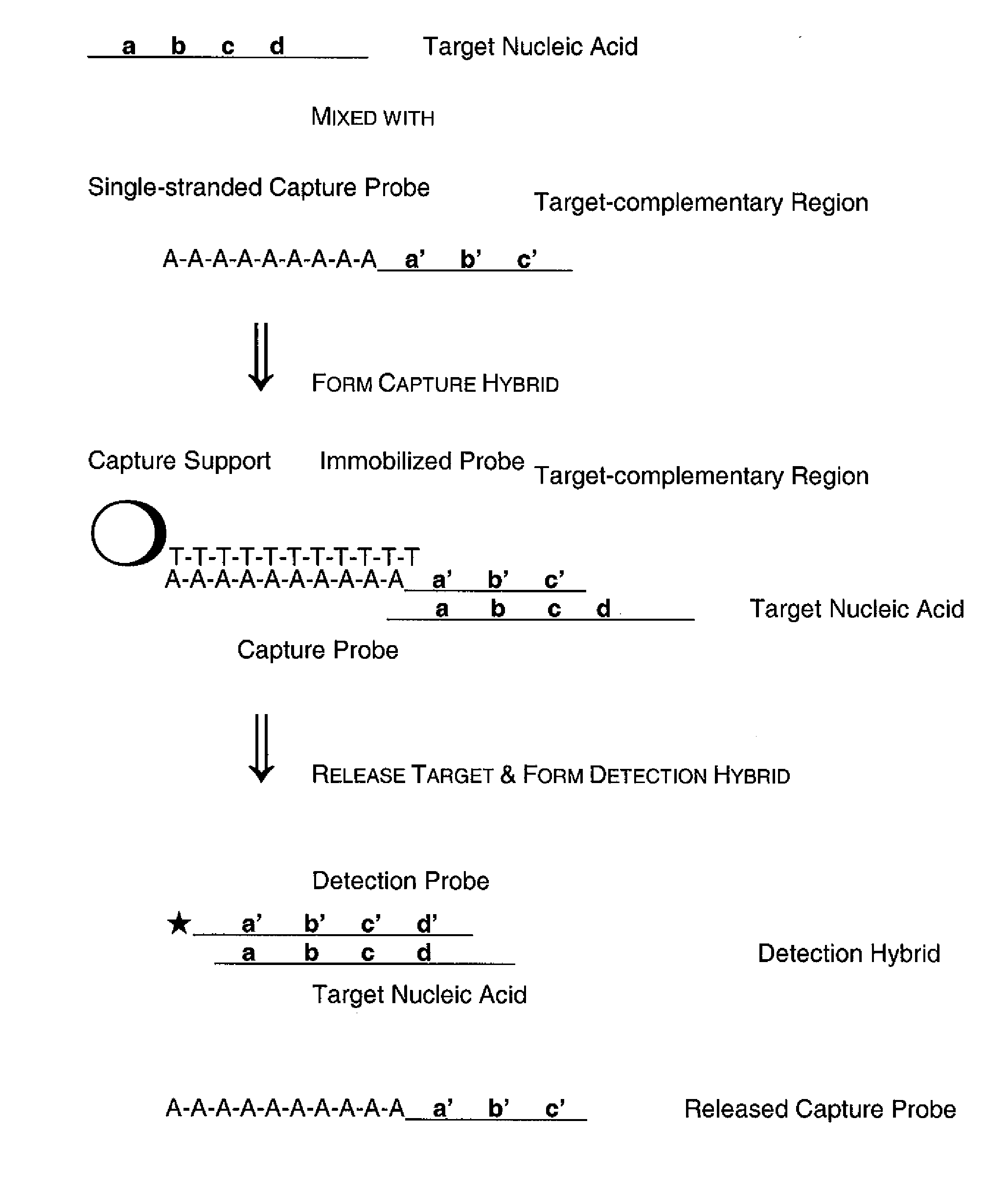
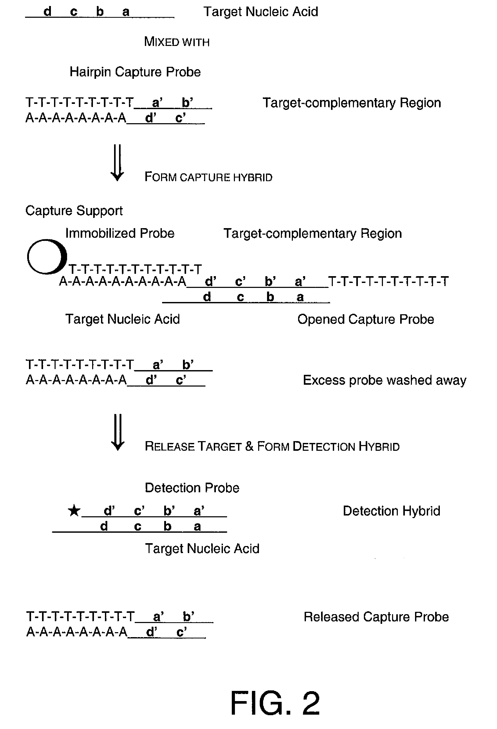
[0050]In these assays, a capture probe capable of forming a hairpin structure under hybridization conditions was used to capture a target nucleic acid from a sample, followed by hybridization of the target nucleic acid with a labeled detection probe and detection of a signal from bound detection probe. The capture probes used in these experiments all contain structural features that allow formation of a hairpin structure: a 5′ region homopolymeric sequence, a 3′ region sequence that was fully or partially complementary to the 5′ region sequence, and a target-complementary sequence flanked by the 5′ and 3′ region sequences. The 5′ and 3′ region sequences form the “stem” portion of the hairpin structure, and the target-complementary sequence forms the “loop” portion of the hairpin structure.

[0051]Three versions of a hairpin capture probe were synthesized and assayed using routine methods to determine the Tm of the stem of the hairpin capture probe....


PUM


Abstract

Methods of the invention separate a target nucleic acid from a sample by using at least one capture probe oligonucleotide that contains a target-complementary region and a member of a specific binding pair that attaches the target nucleic acid to an immobilized probe on a capture support, thus forming a capture hybrid that is separated from other sample components before the target nucleic acid is released from the capture support and hybridized to a detection probe to form a detection hybrid that produces a detectable signal that indicates the presence of the target nucleic acid in the sample. Compositions for practicing the methods of the invention include a capture probe oligonucleotide made up a target-complementary region sequence and a covalently linked capture region sequence that includes a member of a specific binding pair.

Description

RELATED APPLICATION[0001]This application is a continuation of U.S. Ser. No. 11 / 173,915, filed Jun. 29, 2005 and which claims the benefit under 35 U.S.C. 119(e) of provisional application No. 60 / 585,421, filed Jul. 1, 2004. Both are incorporated by reference herein.FIELD OF THE INVENTION[0002]The invention is related to the field of nucleic acid assays, and particularly to detection of a specific target nucleic acid present in a sample, such as small RNA or DNA sequence present in a biological sample.BACKGROUND[0003]Detection of nucleic acids in a sample is useful in diagnostic, therapeutic, forensic, agricultural, food science applications and other areas. Methods of nucleic acid detection include those that use physical separation and detection of a nucleic acid, such as by capturing the nucleic acid in or on a matrix or support and detecting the captured nucleic acids by using a means to visualize the nucleic acid, such as a dye or intercalating agent, or by hybridizing a detecta...

Claims


Application Information

Patent Timeline
Patent Type & Authority Applications(United States)
IPC IPC(8): C12Q1/68G01N33/569C07H21/00G01N33/53
CPCC12Q1/6834C12Q1/6837C12Q2565/525C12Q2565/501C12Q2525/301
Inventor BECKER, MICHAEL M.MAJLESSI, MEHRDAD R.
Owner GEN PROBE INC
Why Eureka
Try Eureka