
Devices and methods for resecting soft tissue

a soft tissue and device technology, applied in the field of medical devices, can solve the problems of complex procedures, inconvenient multi-step process, and large time-consuming, and achieve the effect of preventing unstable fluttering of the shuttle body

Inactive Publication Date: 2013-08-15
MYROMED LLC
View PDF13 Cites 53 Cited by
  • Summary
  • Abstract
  • Description
  • Claims
  • Application Information

AI Technical Summary

Benefits of technology

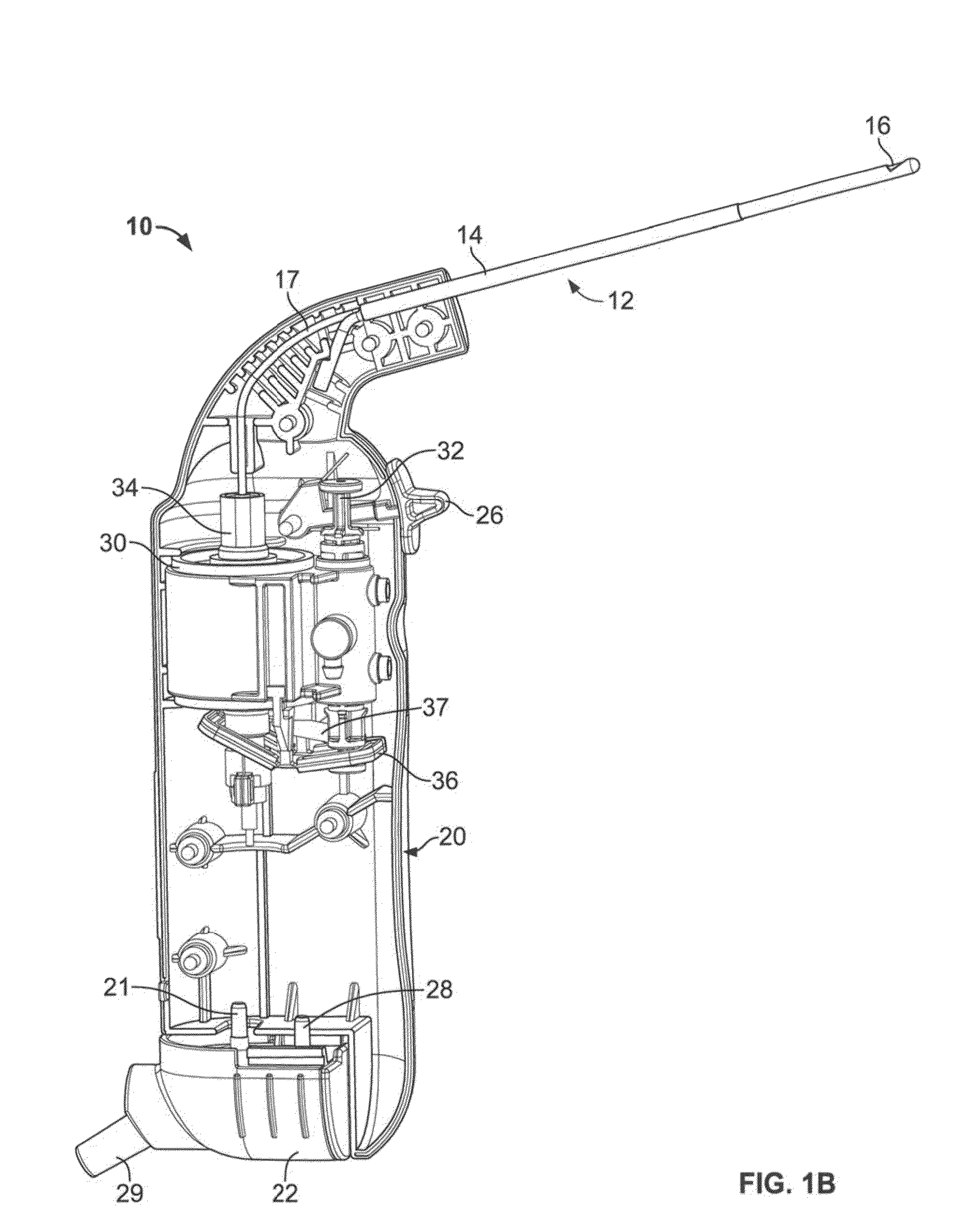
The patent describes a medical device that is powered by a vacuum source. The device has a handle with a linkage support element, and a working end with an operable element that is connected to a mechanism in the handle. When the mechanism is driven by the vacuum source, the operable element moves. The device also has a shuttle body that can move between a forward and return position to switch the fluid path between the chamber and the vacuum source. The linkage connects the drive shaft to the shuttle body to prevent instability in the shuttle body and to support it in the first position before use. The linkage is unsupported by the linkage support element in the second position after use. The technical effects of this invention are a more stable and reliable medical device that can be easily controlled by a vacuum source.

Problems solved by technology

These more complex procedures are typically done in an operating room setting.
The surgical tools are very small and require a significant amount of time to complete the procedure.
A ring curette is capable of cutting tissue and dragging it out of the uterus however, several passes are required to remove the tissue which causes patient discomfort and the device is used without the benefit of visualization.
This multi-step process is frequently inconvenient and time consuming.
The use of multiple instruments for cutting and extracting tissue increases cost due to the expense of purchasing separate tools and increased procedure time.
Furthermore, excised tissue may be friable which makes it difficult to remove through a cannula because it breaks into pieces when grasped and / or when it comes into contact with the cannula.
In addition to being time consuming and tedious, some of the tissue may be “lost” and become a nidus for infection.

Method used


Image

  • Devices and methods for resecting soft tissue
  • Devices and methods for resecting soft tissue
  • Devices and methods for resecting soft tissue

Examples


Embodiment Construction

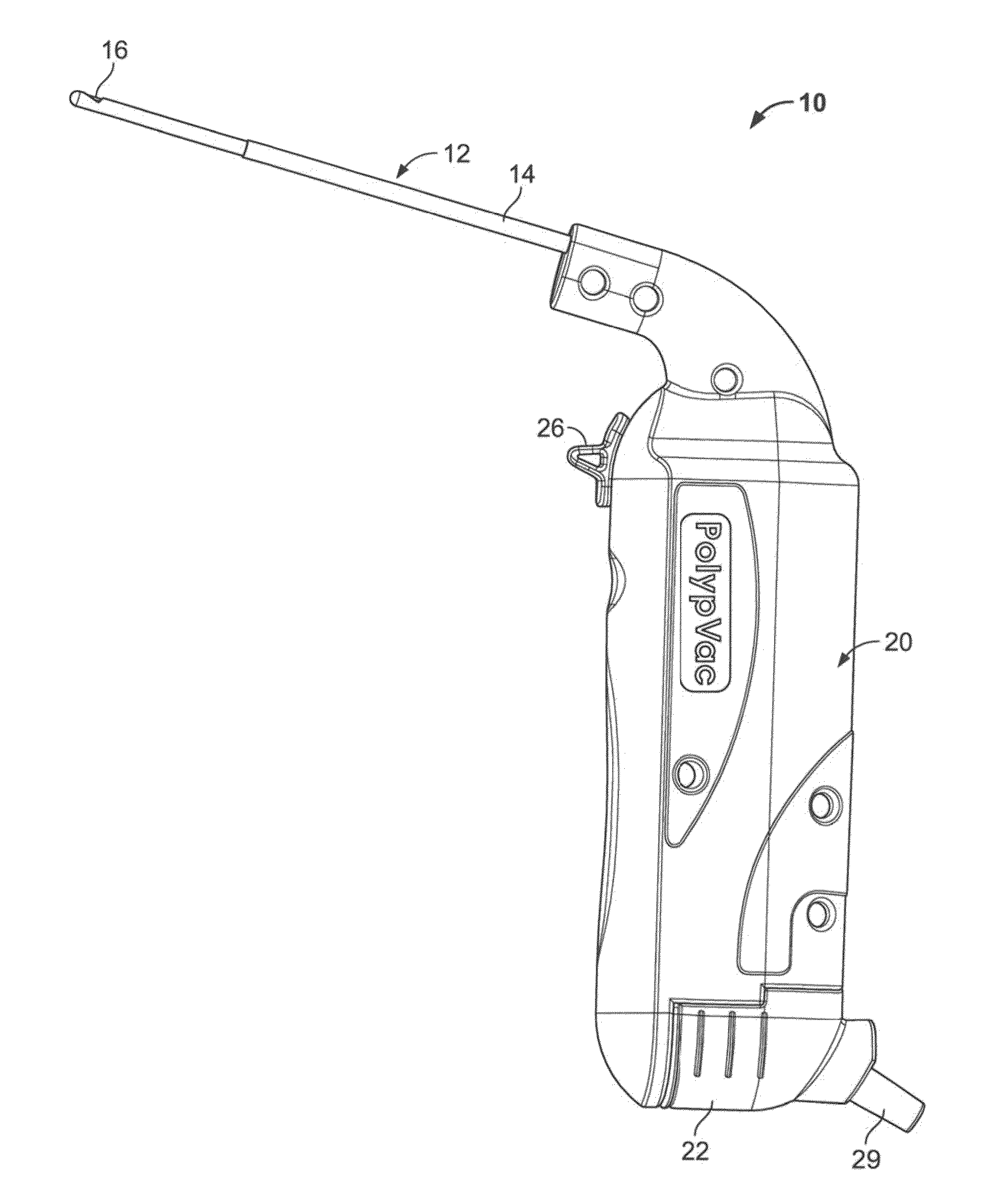
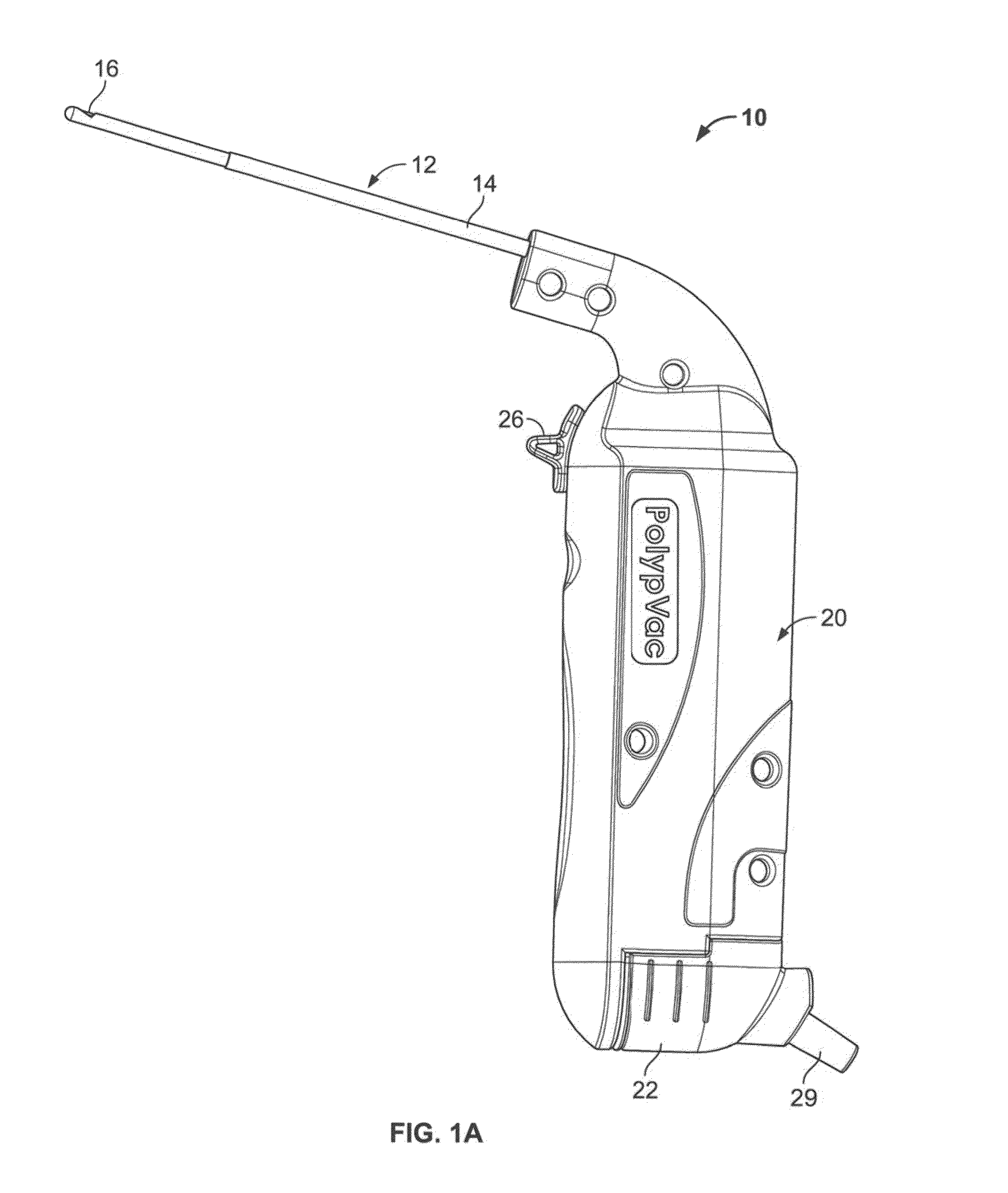
[0069]Variations of the devices are best understood from the detailed description when read in conjunction with the accompanying drawings. It is emphasized that, according to common practice, the various features of the drawings may not be to-scale. On the contrary, the dimensions of the various features may be arbitrarily expanded or reduced for clarity. The drawings are taken for illustrative purposes only and are not intended to define or limit the scope of the claims to that which is shown.

[0070]Various medical devices, including various cutting devices and methods for cutting, resecting, incising or excising tissue are described herein. In certain variations a medical device may include a mechanism or motor driven or powered by a variety of different power sources, e.g., suction from a vacuum source, pneumatic, fluid pressure (e.g. hydraulic), compressed air, battery power or electrical power or gas power or any combination thereof. The mechanism or motor may create a reciproca...


PUM


Abstract

The present devices and methods relate generally to medical devices and methods for cutting, resecting, excising and / or evacuating tissue from various regions of a patient's body. In particular, devices and methods for cutting and / or removing various tissues, e.g., polyps or endometrium, from a uterus or uterine space are provided. Devices and methods for cutting and / or removing various tissues from the human body, such as during laparoscopic or thoracoscopic surgery, are also provided.

Description

CROSS-REFERENCE TO RELATED APPLICATIONS[0001]This application claims the benefit of priority to U.S. Provisional Patent Application No. 61 / 597,642 filed Feb. 10, 2012, the contents of which are incorporated herein by reference in their entirety.FIELD OF THE INVENTION[0002]The present devices and methods relate generally to medical devices and methods for performing work on tissue, for example, cutting and / or evacuating tissue from various regions of a patient's body.BACKGROUND[0003]A hysteroscopy is a procedure for women that may be used to evaluate and treat a variety of abnormal conditions in the uterus. Diagnostic hysteroscopy may help diagnose problems causing heavy bleeding such as fibroids and polyps. Hysteroscopy for the purpose of diagnostics and some simple operative procedures can be performed in a doctor's office under local anesthesia. Operative hysteroscopy may be used to remove polyps or fibroids, cut adhesions, correct uterine abnormalities, manage blocked fallopian t...

Claims


Application Information

Patent Timeline
IPC IPC(8): A61B17/32A61M13/00
CPCA61M1/0058A61B17/320708A61B17/42A61M13/003A61M13/00A61B2017/320028A61B2017/4216A61B17/32002A61B2017/320008A61B17/320016A61B17/3205A61B2017/320024A61B2017/32004A61B2017/320064A61B2217/005A61B2217/007
Inventor DUBOIS, BRIAN R.NIELSEN, JAMES T.GORDON, ALEXANDER
Owner MYROMED LLC
Learn More