De Novo Anembryonic Trophoblast Vesicles and Methods of Making and Using Them
a technology of embryonic trophoblasts and vesicles, applied in the field of biotechnology, can solve the problems of inability to study the complex cell-to-cell interaction of neither model, and the technique does not allow the study of cell-to-cell interactions, and achieve the effects of improving the ability of scientists, improving efficiency, and improving understanding
- Summary
- Abstract
- Description
- Claims
- Application Information
AI Technical Summary
Benefits of technology
Problems solved by technology
Method used
Image
Examples
example 1
Trophoblast Cells Form Trophoblast Vesicles in a Nonadhesive Micro-Mold Bioreactor
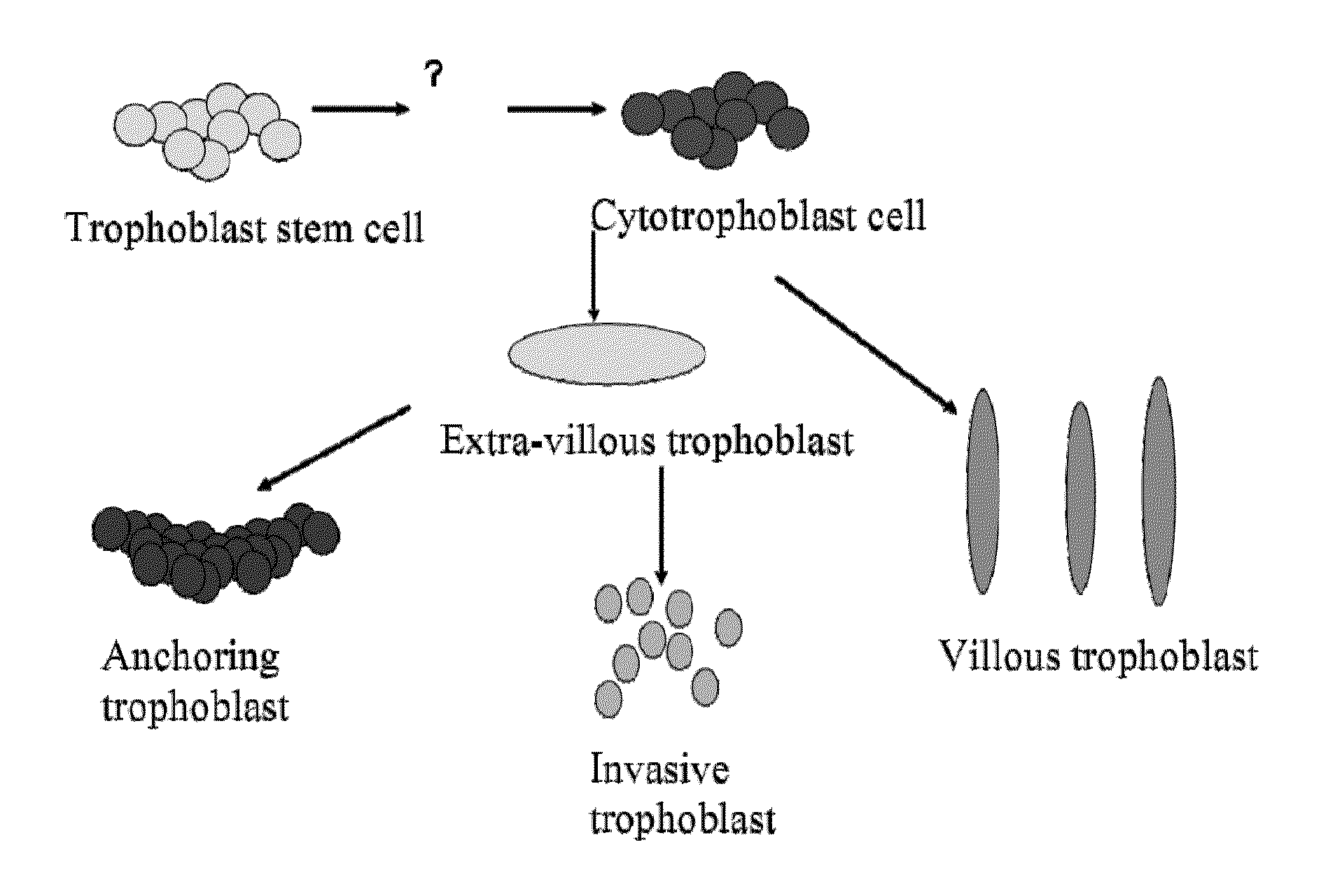
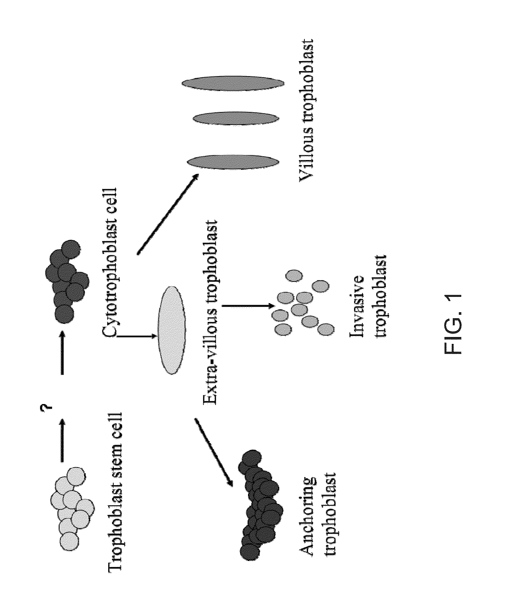
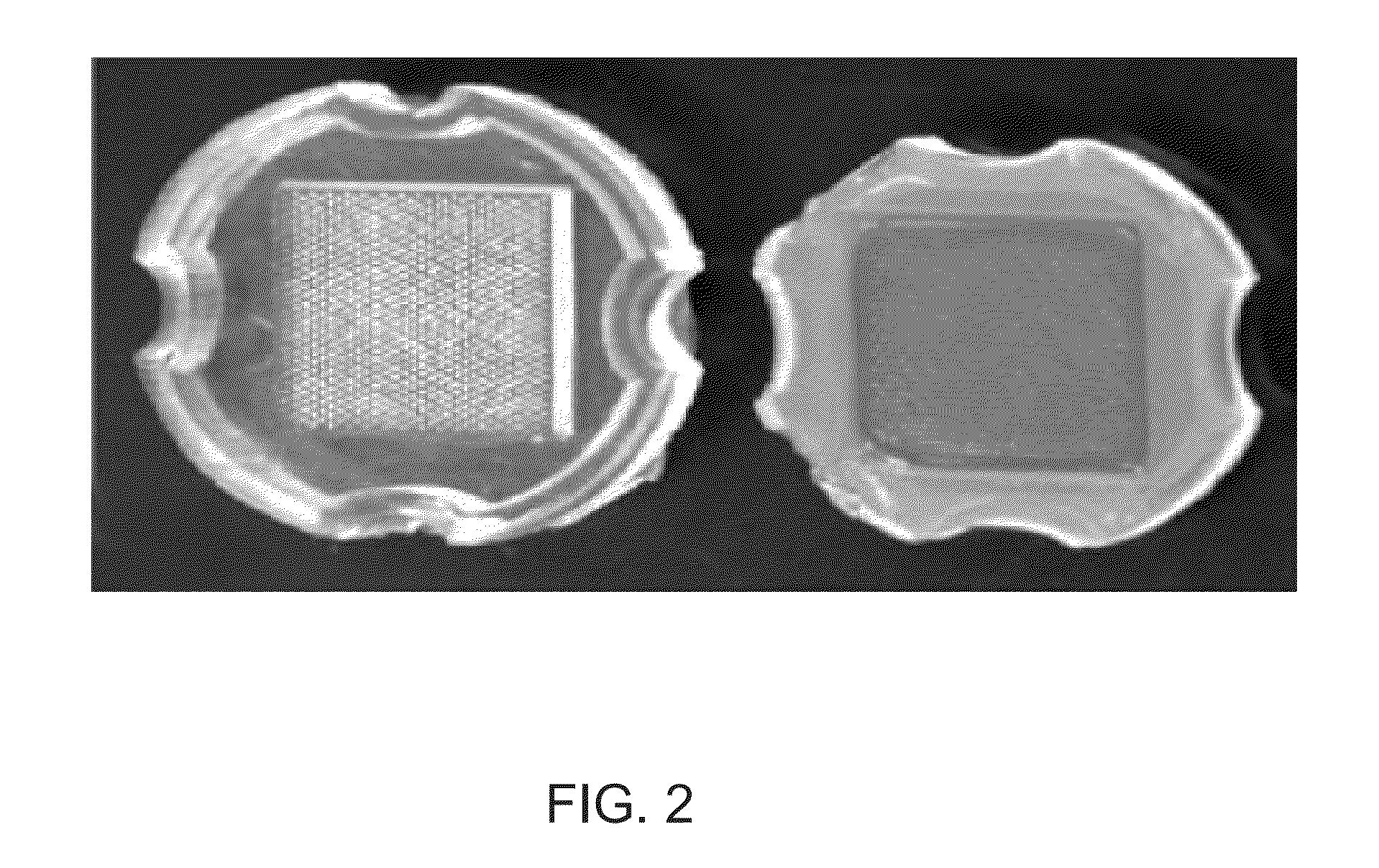
[0033]Human immortalized trophoblast cells were seeded into nonadhesive micro-mold bioreactors containing 822 cylindrical recesses 200 μm in diameter. Each recess was seeded with 800,000 cells. The cells settled into the wells within hours of seeding. Within three days, the cells formed uniform sized spheroids approximately 150 μm in diameter (see FIG. 4) Determination of the three-dimensional spheroid morphology and was performed on days 7 and 10 after seeding. The spheroids were removed from the molds, fixed and sectioned. Surprisingly, the spheroids contained a hollow center with a cellular rim 12.3 μm (±1 μm) in thickness (see FIG. 4). The morphology did not change significantly between days 7 and 10; the thickness of the rim and the diameter of the center remained constant. The consistent hollow shape of the sphere was confirmed in live cells with confocal imaging after DAPI staining (see FIGS. 4-...
example 2
Trophoblast Cells in Vesicles are Highly Active and Form Tight-Junctions
[0034]Electron microscopy was performed to further demonstrate cellular morphology and examine cell-to-cell interactions. Close inspection of individual cells at high power demonstrated that the cytoplasm was replete with cellular organelles including large numbers of mitochondria, endoplasmic reticulum and golgi. This confirms that the cells are alive and, more importantly, metabolically active. Mitotic bodies can be identified illustrating that the cells in the rim are proliferating. Apoptotic bodies can be also identified throughout the cellular rim adjacent to mitotic cells. Numerous tight-junctions were identified among the viable cells supporting the notion that cell-to-cell communication is occurring (see FIGS. 7-8). This formation of tight junctions is a hallmark of in vivo trophoblast vesicle formation.
example 3
Trophoblast Vesicles are Highly Adherent
[0035]Spheroids removed from the molds adhere to the cell culture dishes within two hours. These cells will proliferate across the surface of the plate and form confluent monolayers. This proliferative action is morphologically similar to a blastocyst attached to a cell culture dish. Upon reseeding into the aggregation device, the cells in monolayer will re-form spheroids. There are no obvious morphologic differences among the spheroids formed by multiple reseedings. These data indicate that the trophoblast cells are forming biologically active vesicles. These de novo vesicles may be employed in implantation, using various known in vitro and in vivo models of tissue function and implantation.
PUM
| Property | Measurement | Unit |
|---|---|---|
| depth | aaaaa | aaaaa |
| depth | aaaaa | aaaaa |
| width | aaaaa | aaaaa |
Abstract
Description
Claims
Application Information
 - R&D
- Intellectual Property
- Life Sciences
- Materials
- Tech Scout
- Unparalleled Data Quality
- Higher Quality Content
- 60% Fewer Hallucinations
Browse by: Latest US Patents, China's latest patents, Technical Efficacy Thesaurus, Application Domain, Technology Topic, Popular Technical Reports.
© 2025 PatSnap. All rights reserved.Legal|Privacy policy|Modern Slavery Act Transparency Statement|Sitemap|About US| Contact US: help@patsnap.com


