System and method for remote control of a microscope

a technology of remote control and microscope, which is applied in the field of remote control of automated microscope, can solve the problems of not providing for such easy collaboration and possible catastrophic failur

Inactive Publication Date: 2014-02-20
IKONISYS INC
View PDF9 Cites 14 Cited by
  • Summary
  • Abstract
  • Description
  • Claims
  • Application Information

AI Technical Summary

Benefits of technology

[0012]Experts may also differ on what components of an image may be significant. Thus, one expert may see a possible aberration from normalcy in a portion of an image, wherein another may not. When questions arise today, new samples are often collected and new images taken to determine whether a hypothesized aberration truly exists. There is not provided a mechanism wherein one or more investigators seeking the microscopic services of a remote location can request in real time a closer view of a specimen locus based upon a predefined image resolution. In an embodiment, the present invention overcomes such problem by allowing one or more investigator to review the microscopic examination of the specimen in real time, and to request through remote control of the microscope for more detailed examination of one or more locus of the specimen, altering parameters of the scope such as magnification and filtering.
[0013]It may be advantageous after receiving a microscopic image and making a diagnosis there from, to be able to revisit the slide in the future. In fact, regulations may require that a specimen sample on a microscope slide be archived in order to allow post-diagnosis review of a diagnostic decision. There can be advantage in having a central microscopy location which is equipped to maintain such slides in a storage environment that protects the integrity of the specimen on the slide to the greatest extent. Retrieving a single slide from many may however be a difficult task. In an embodiment, there is therefore provided a system for cataloguing a particular slide along with a particular automated microscope cassette and the position of such slide in the cassette. In such embodiment the automated microscope uses such information to automatically select the remotely requested slide from the appropriate cassette and to provide image access to the slide to the remote person seeking the same. Optionally, data storage may be associated with the slide and / or automated microscope that can be accessed by the automated microscope to concomitantly provide the image data originally associated with the slide when first probed (and / or the scanning parameter data on how such image was constructed). By comparing such original image data to any image data later obtained on the specimen, a determination of the degree of degradation of the specimen over time, if any, may be made. Retrieval of the slide may allow one to visit other areas of the slide / sample that were not part of the original scanning results, or to more closely review a portion of sample on the slide. An embodiment automated microscopy systems provides the ability to revisit a previously scanned area of a slide / sample by automatically loading and positioning the slide to the area of interest.

Problems solved by technology

When laboratory technician performs original analysis on a microscope specimen in a location remote to the person seeking information about the specimen, such as a clinician, collaboration by multiple persons interested in the microscopic analysis, such as other engineers, clinicians, or researchers, entails a time consuming post examination transmission of multiple copies of an image and exchange of views by each of the reviewers.
Likewise, structural engineers facing a possible catastrophic failure of a structure may need to understand immediately the condition of structure and may need to consult others on whether the structure should be closed.
Present systems, however, do not provide for such easy collaborations unless everyone is located in the same location and has ready access to the microscopic image.

Method used


Image

  • System and method for remote control of a microscope
  • System and method for remote control of a microscope
  • System and method for remote control of a microscope

Examples


Embodiment Construction

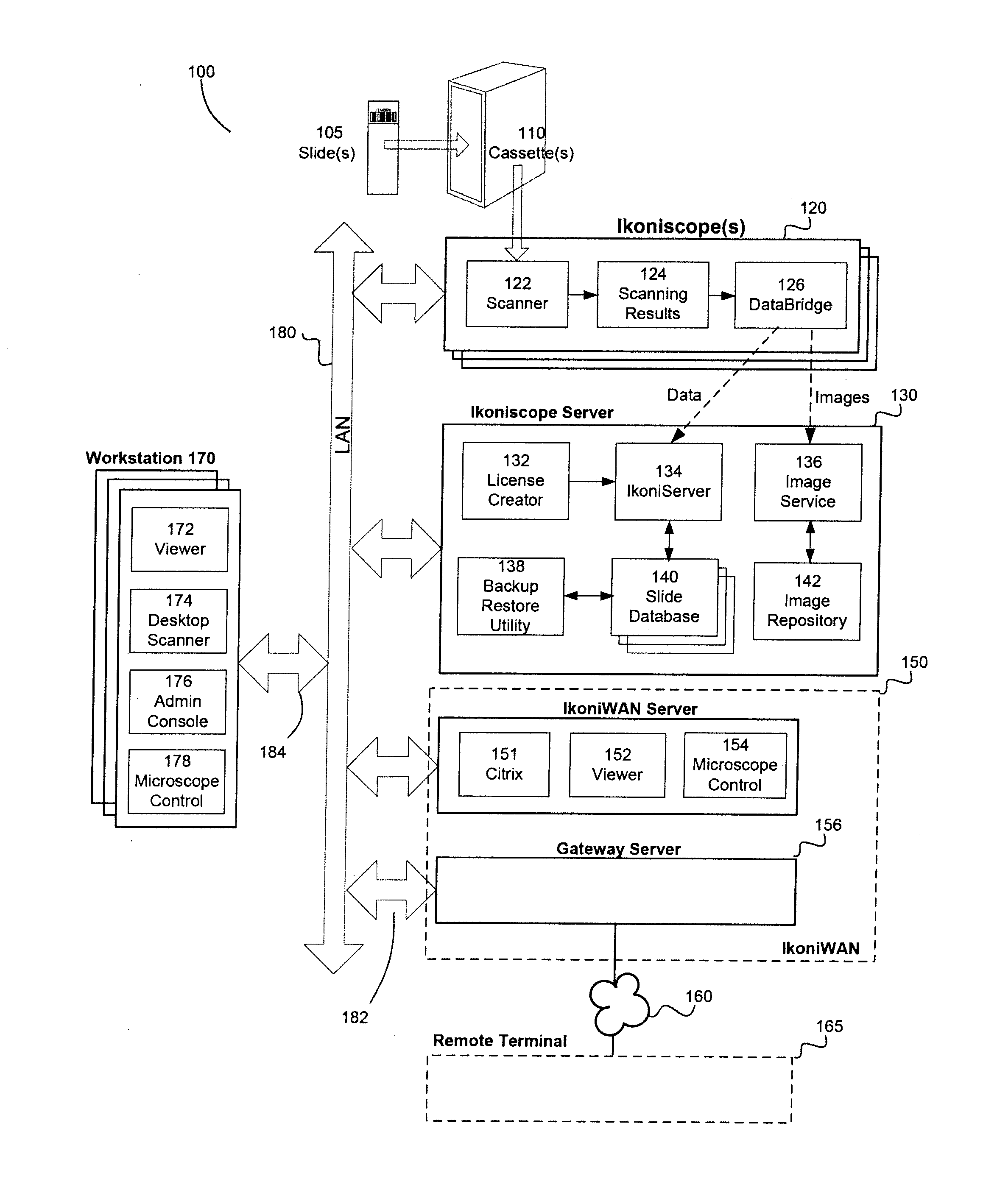
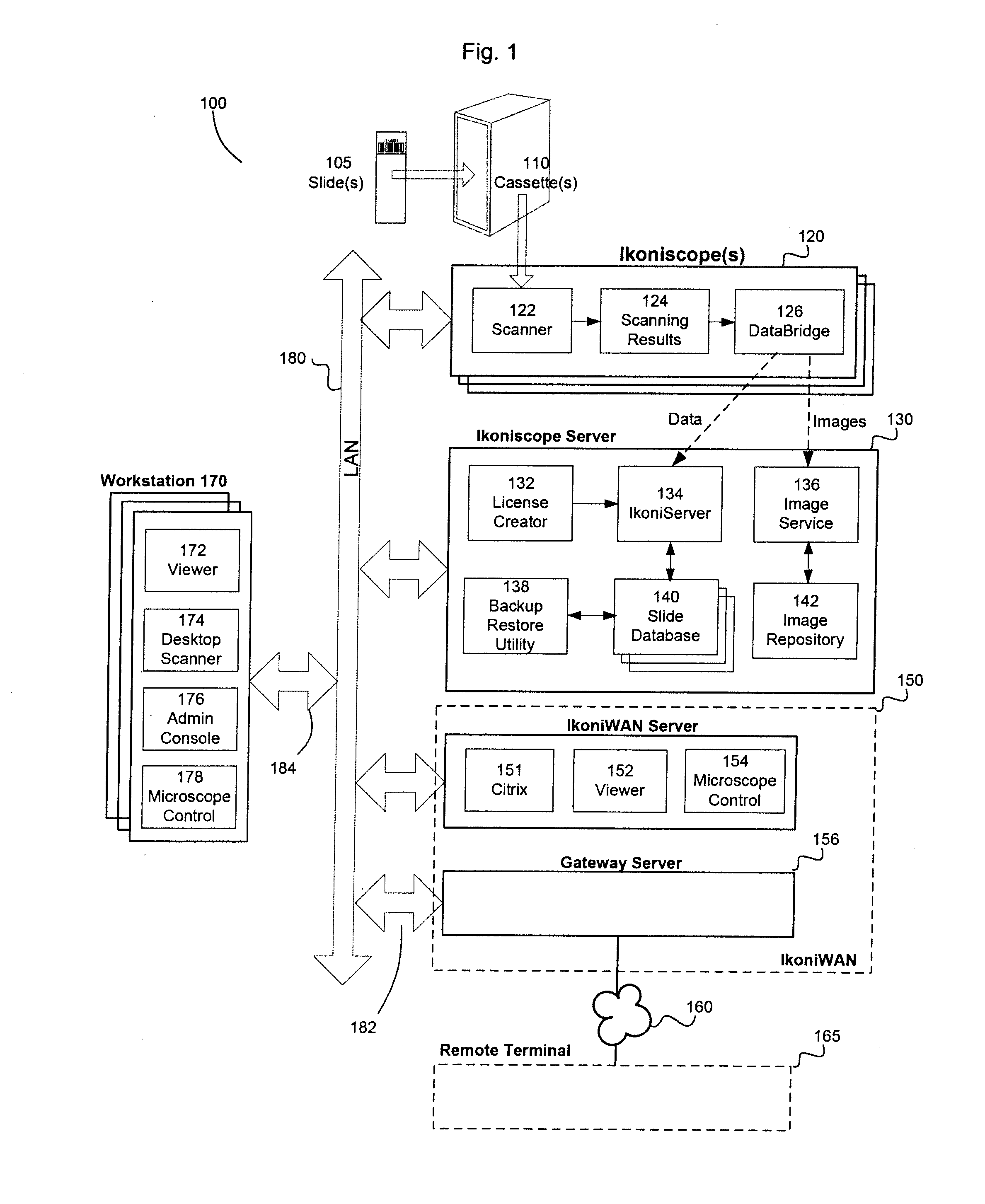
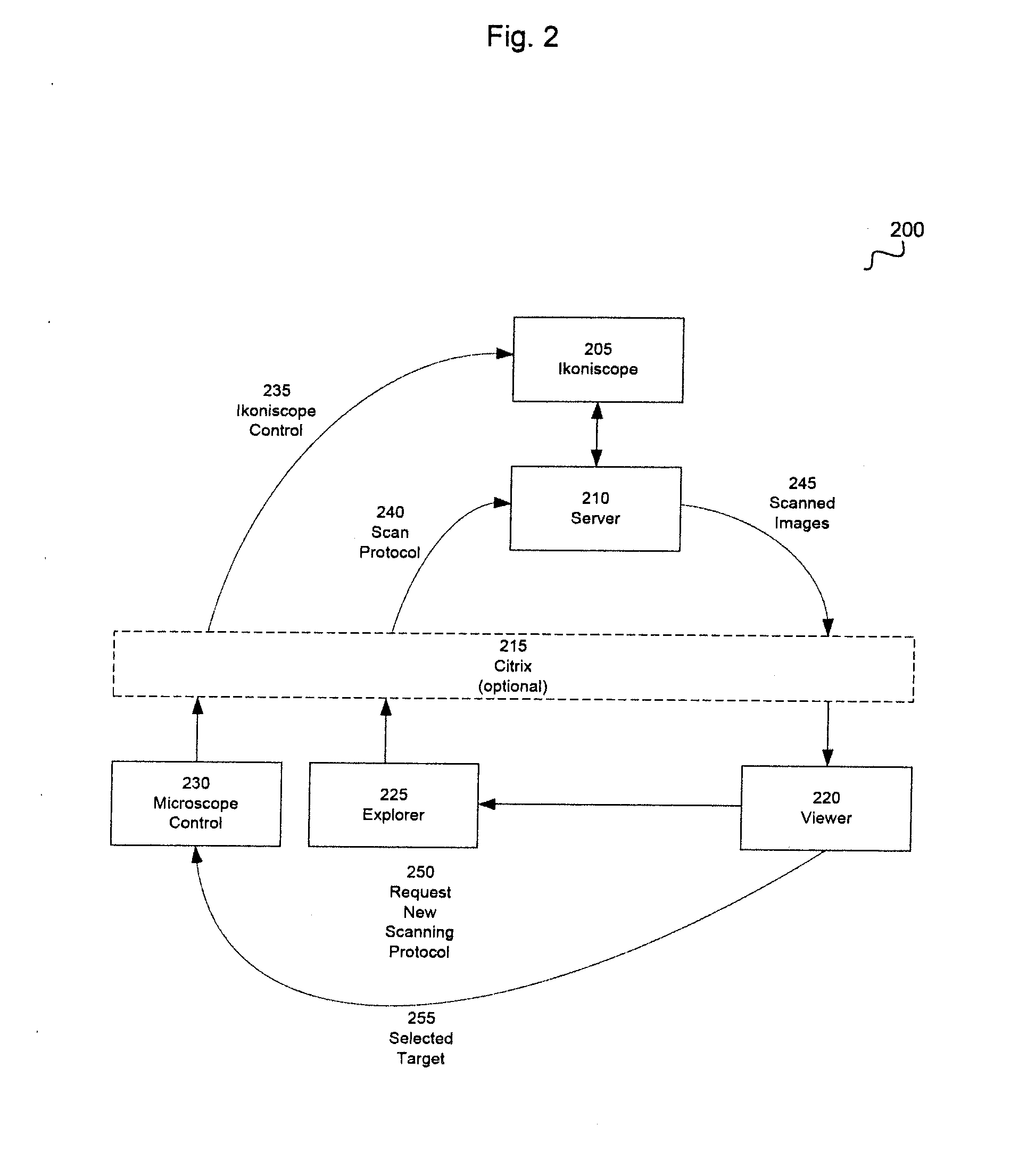
[0031]The present invention generally relates to a system and method for remote control of an automated microscope.

[0032]The remote system and method features are a sub-system, integrated into an automated microscope system, such as can be found in the Ikonisys, Inc., Ikonisoft software system and Ikoniscope automated microscope. The system and methods herein provide the capability to remotely control an automated microscope system and capture and transmit imagery data to one or more remote workstation in real-time.

[0033]The following definitions will be found useful in describing the system and method of the present invention:

[0034]Cassette: A slide container capable of holding a plurality of slides in a non-contacting fixed position.

[0035]Channel: A combination of excitation filter, dichroic mirror, and emission filter utilized to produce fluorescent image at a given magnification.

[0036]DAPI: 4′6-diamindino-2-phenylindole-2HCl, a fluorescent probe for DNA used for nucleus visualiz...


PUM


Abstract

A system and method for remote control of an automated microscope via a widely distributed network.

Description

RELATED APPLICATIONS[0001]This application is a continuation of the patent application Ser. No. 12 / 817,132 filed Jun. 16, 2010. U.S. Provisional Application 61 / 187,422 filed Jun. 16, 2009; and U.S. Provisional Application 61 / 333,892 filed May 12, 2010. All related applications are incorporated herein by reference in their entirety.BACKGROUND OF THE INVENTION[0002]1. Field of the Invention[0003]The present invention generally relates to a remotely controlled automated microscope.[0004]2. Description of the Related Art[0005]In a conventional optical microscope, a single objective lens is used to focus on one location of a specimen carried by a microscopic slide and to acquire an image of the location. Dimensions of the image depend on the magnification and the numerical aperture of the objective lens. The image is viewed through an ocular lens or acquired through a camera. The specimen is moved and the same image-viewing process is repeated at a new location. When using these manual m...

Claims


Application Information

Patent Timeline
Patent Type & Authority Applications(United States)
IPC IPC(8): G02B21/00H04N7/18
CPCH04N7/18G02B21/008G02B21/368H04N7/181G02B21/365G16H10/40Y02A90/10H04N23/66G02B21/34
Inventor TAFAS, TRIANTAFYLLOS P.THOMAS, MICHAELKIM, YOUNGMINBORGERDING, ROBERT H.WESTPHAL, FRED C.
Owner IKONISYS INC
Why Eureka
Try Eureka