Systems and methods for interactive magnetic resonance imaging

a magnetic resonance imaging and interactive technology, applied in the field of interactive magnetic resonance imaging, can solve the problems of increasing procedural complexity, unable to provide reliable images for use in assessing certain pathological conditions of patients, and difficulty in insertion and navigation of interventional devices within different branches of the vascular system,

Inactive Publication Date: 2014-12-11
GENERAL ELECTRIC CO
View PDF9 Cites 139 Cited by
  • Summary
  • Abstract
  • Description
  • Claims
  • Application Information

AI Technical Summary

Problems solved by technology

X-ray and CT images, however, visualize soft tissues poorly, and thus, may not provide reliable images for use in assessing certain pathological conditions of the patient.
Moreover, X-ray and CT imaging exposes the patient and an operator to ionizing radiation that may increase with procedural complexity.
Insertion as well as navigation of the interventional device within different branches of a vascular system, however, is a challenging procedure.
Further, identification of the desired anatomical features of interest and / or an accurate imaging plane may be confounded due to noise and poor contrast between target structures and surrounding tissues.
Additionally, dynamic prescription of scanning protocols and accurate selection of scanning parameters suitable for a specified imaging task are time consuming and often dependent on the operator's experience and skill.
Particularly, in absence of relevant experience, navigation to a desired region, selection of appropriate scanning protocols and / or landmark recognition from a novice operator may often result in inconsistent and / or inaccurate imaging.
Such inaccurate estimations or inadequate coverage of clinically relevant parameters such as a location of a lesion derived from images reconstructed using erroneous configurations may lead to incorrect diagnosis, which in turn, may adversely affect patient health.
Additionally, inadequate coverage of the desired ROI may necessitate additional data acquisition, which further increases exam duration, while also adding to patient discomfort.
Moreover, even when experienced operators are available, manual intervention based imaging and navigation may become a bottleneck for large-scale deployment of image analysis techniques.

Method used


Image

  • Systems and methods for interactive magnetic resonance imaging
  • Systems and methods for interactive magnetic resonance imaging
  • Systems and methods for interactive magnetic resonance imaging

Examples


Embodiment Construction

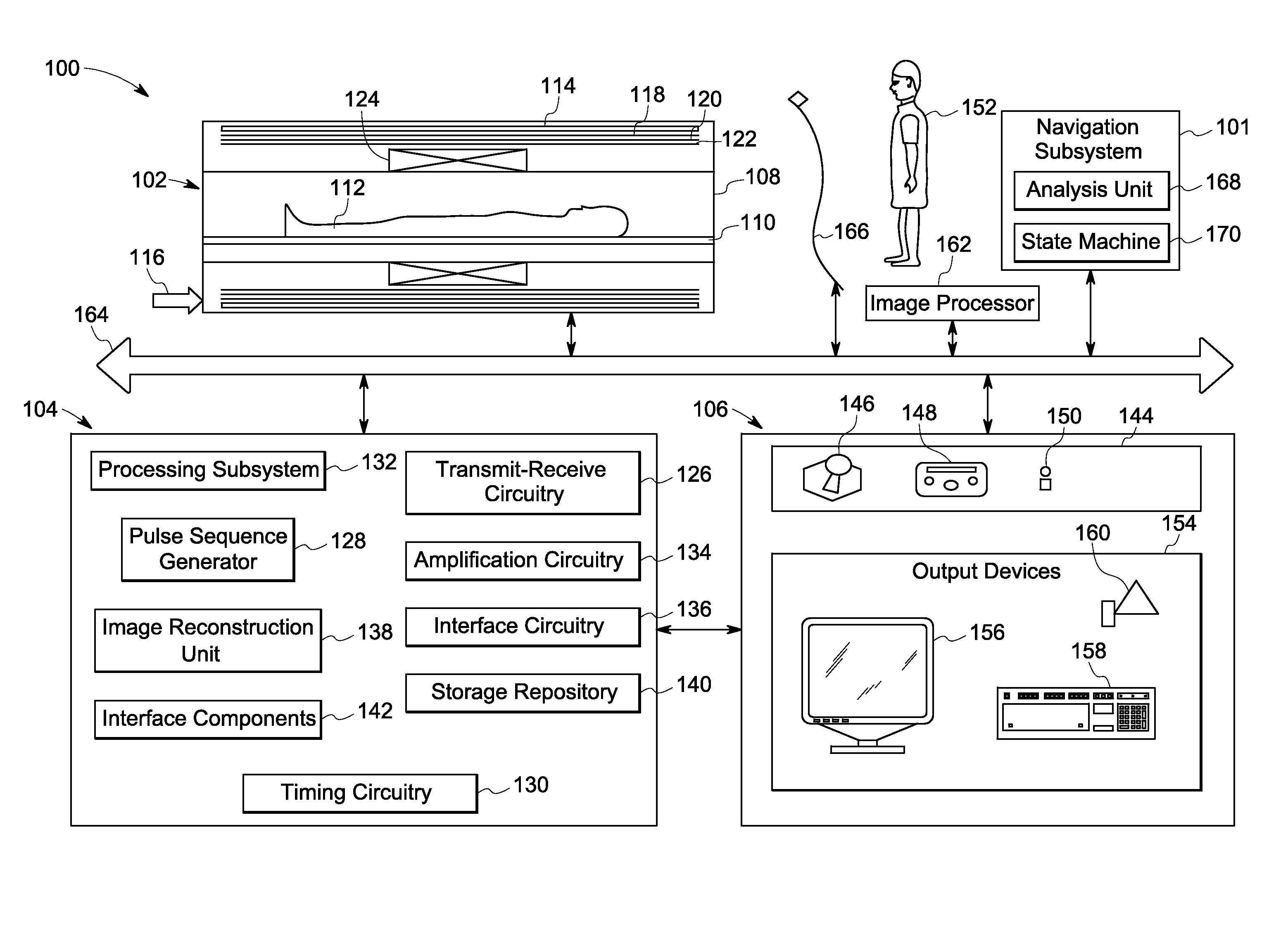
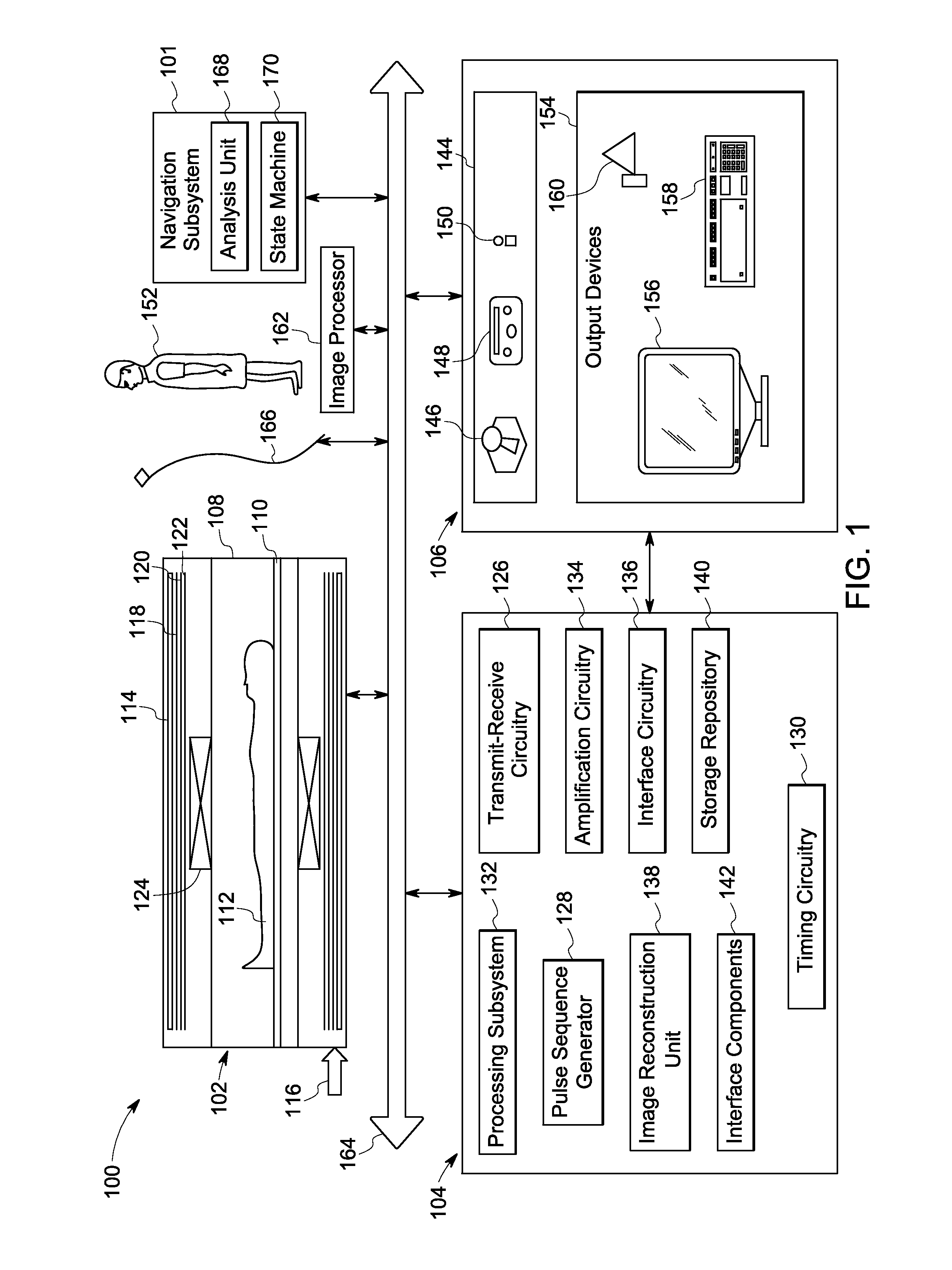
[0011]The following description presents exemplary systems and methods for interactive navigation of a human body during diagnostic imaging. Particularly, embodiments illustrated hereinafter disclose an anatomy-aware MRI system that may be configured to allow interactive and real-time navigation of the human body by even inexperienced operators. Embodiments of the anatomy-aware MRI system may also be configured to perform a variety of imaging tasks, such as imaging a target VOI relative to a current field of view (FOV), labeling features of interest during imaging, and tracking an interventional device based on interactive input received from an operator in real-time.

[0012]Although exemplary embodiments of the present systems and methods are described in the context of magnetic resonance (MR) imaging, it will be appreciated that use of embodiments of the present systems and methods in various other imaging applications and systems is also contemplated. For example, embodiments of th...


PUM


Abstract

Embodiments of a method, a system, and a non-transitory computer readable medium for use in interactive magnetic resonance imaging are presented. An initial region of interest of a subject is scanned to acquire imaging data using an initial imaging protocol. Anatomical labeling information corresponding to a plurality of regions corresponding to the subject may be determined based on the acquired data and/or previously available information. Particularly, determining the anatomical labeling information may include identifying one or more features of interest corresponding to the initial region of interest. Further, input from an operator corresponding to a desired imaging task may be received interactively. The imaging protocol may be updated in real-time by selectively configuring one or more imaging parameters that optimize implementation of the desired imaging task based on the determined anatomical labeling information. A subsequent scan may be performed using the updated imaging protocol for completing the desired imaging task.

Description

BACKGROUND[0001]Clinical diagnosis and treatment often rely on image-derived parameters corresponding to a region of interest (ROI) of a patient for managing a plurality of life-threatening medical conditions. Accordingly, various medical procedures employ imaging modalities such as X-ray fluoroscopy, computed tomography (CT), and magnetic resonance imaging (MRI) for generating high-fidelity images that aid in diagnoses and / or guidance of surgical or interventional devices in real-time. Typically, these imaging modalities employ specific scan configurations that allow acquisition and reconstruction of imaging data from a desired ROI of the patient.[0002]X-ray and CT images, however, visualize soft tissues poorly, and thus, may not provide reliable images for use in assessing certain pathological conditions of the patient. Moreover, X-ray and CT imaging exposes the patient and an operator to ionizing radiation that may increase with procedural complexity. Accordingly, certain medical...

Claims


Application Information

Patent Timeline
Patent Type & Authority Applications(United States)
IPC IPC(8): A61B5/00A61B5/055
CPCA61B5/748A61B5/055A61B5/749A61B5/743A61B5/0037G01R33/543G01R33/546A61B5/318
Inventor DARROW, ROBERT DAVIDVAIDYA, VIVEK PRABHAKAR
Owner GENERAL ELECTRIC CO
Why Eureka
Try Eureka