Eureka AIR delivers breakthrough ideas for toughest innovation challenges, trusted by R&D personnel around the world.

Disposable electrosurgical probe and kit and method of using

Active Publication Date: 2015-11-19
ENDOMEDICAL CONCEPTS
View PDF4 Cites 0 Cited by
  • Summary
  • Abstract
  • Description
  • Claims
  • Application Information

AI Technical Summary

Benefits of technology

The invention offers a disposable electrosurgical probe as a kit, with all components (including the working element, sheath, core member, and electrodes) being disposable. This provides a more efficient and cost-effective solution for performing electrosurgical procedures.

Problems solved by technology

As such, electrosurgical probes typically have high initial purchase costs.
The distal end of the sheath is often equipped with a plastic tip that becomes damaged over time, in some cases after a single use, as a result of the high RF current levels, necessitating that the sheath undergo an expensive and time-consuming repair.
Sterilization can be a complicated process, particularly in view of the internal channels within the sheath that accommodate a light source, telescope, and / or cooling flow stopcocks and channels.
If any of the components are unavailable or the wrong size, the procedure cannot go forward.
However, at times when case loads are high, a physician may be forced to either wait for another unit to be sterilized or cancel the surgery.

Method used


Image

  • Disposable electrosurgical probe and kit and method of using
  • Disposable electrosurgical probe and kit and method of using
  • Disposable electrosurgical probe and kit and method of using

Examples


Embodiment Construction

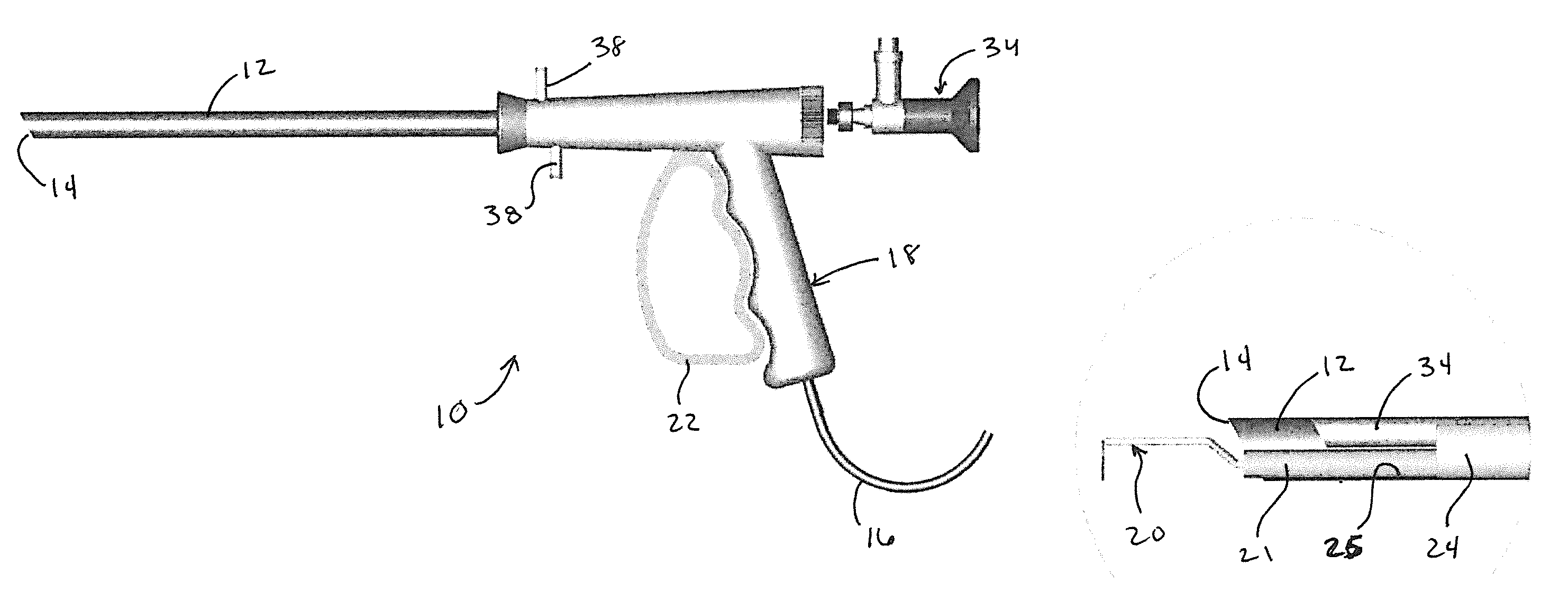
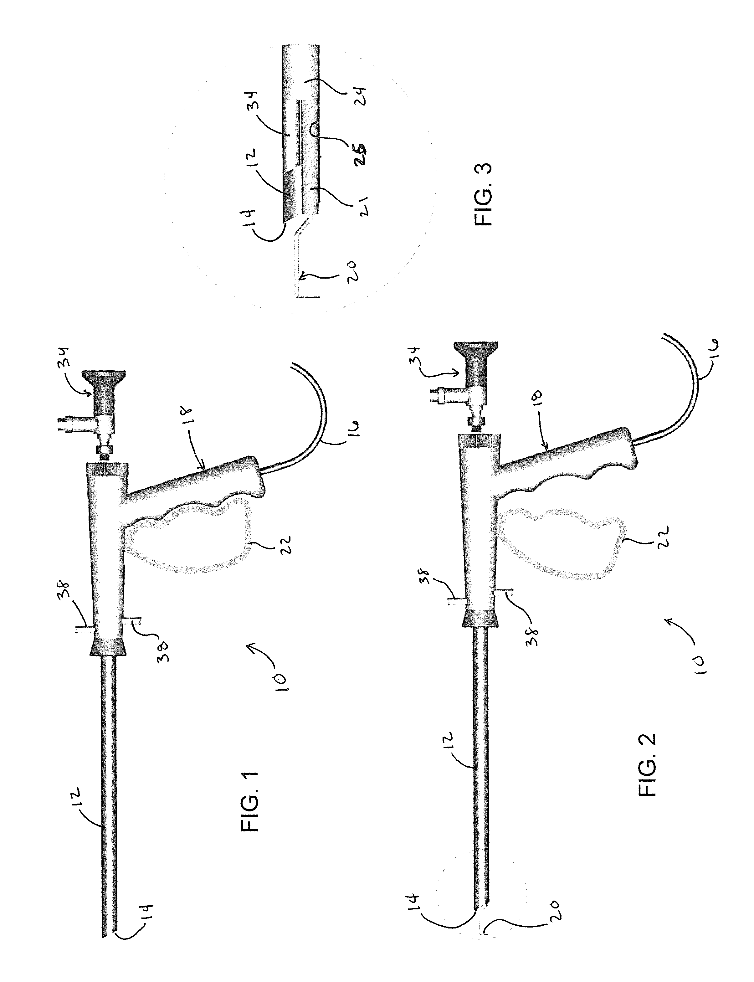
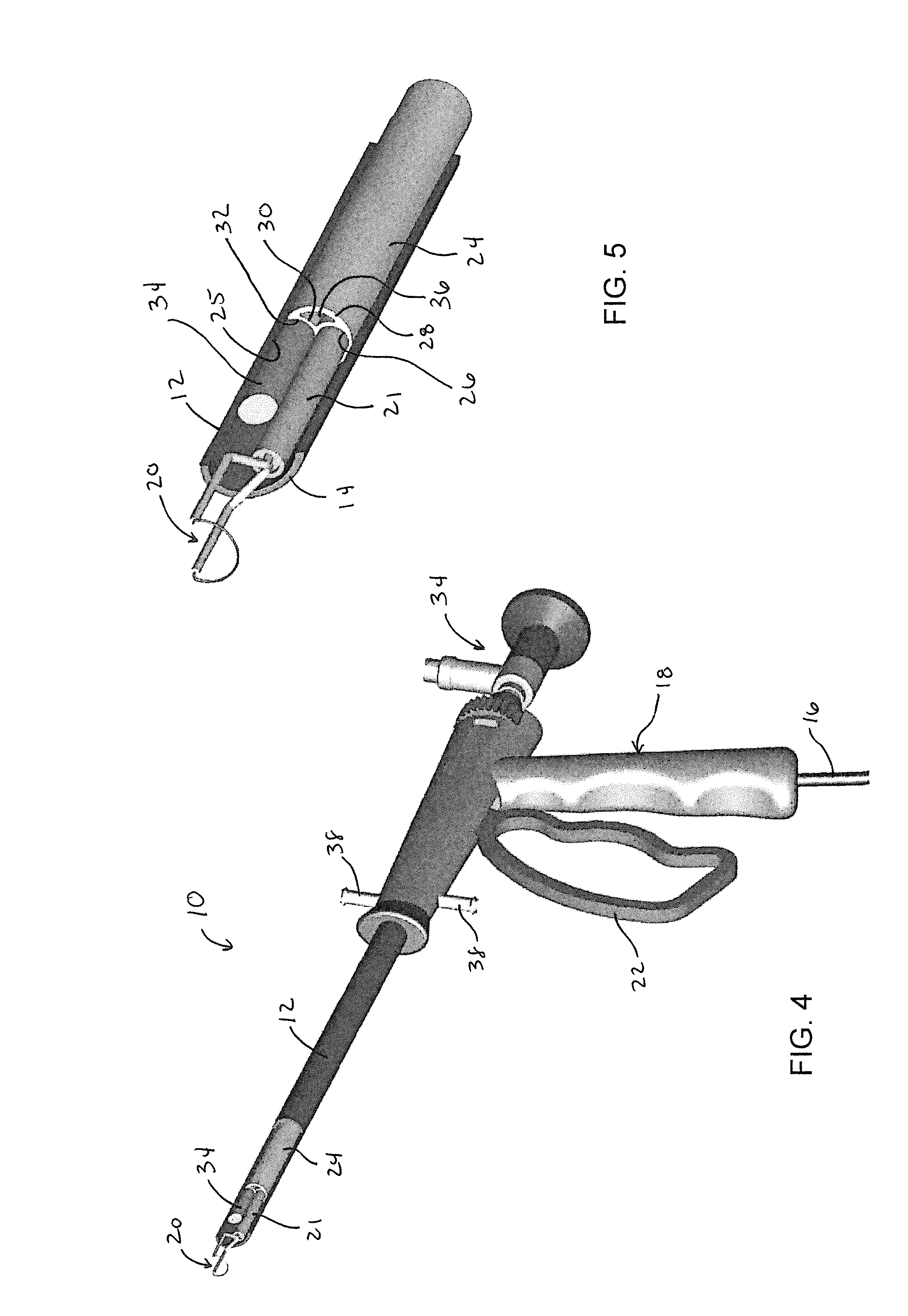
[0018]FIGS. 1 through 5 depict an electrosurgical probe 10 in accordance with a nonlimiting embodiment of the present invention. The drawings depict the probe 10 as a resectoscope, though other electrosurgical probes capable of use in a wide variety of procedures are also within the scope of the invention. The probe 10 is represented as including a sheath 12 through which conductors can be routed to one or more electrodes disposed at a distal end 14 of the sheath 12. The conductors carry a current, preferably an RF current, generated by an electrosurgical generator (not shown) that is connected to the probe 10 via a power cord 16 extending from a working element 18 of the probe 10. The probe 10 can be a monopolar or bipolar RF electrosurgical probe. FIGS. 1 through 5 depict a single electrode 20 configured as a cutting loop (wire), though other electrode configurations are possible and within the scope of this invention, for example, such well-known types as ball tip, disk, roller t...


PUM


Abstract

Disposable electrosurgical probes for treating tissue, and surgical procedures that make use of such probes. Such a probe includes a working element, an elongate sheath secured to the working element, and a core member within the sheath. At least one active electrode and conductor are disposed in a first of a plurality of internal longitudinal channels within the core member. The electrode is adapted to extend from a distal end of the sheath and configured to perform cutting, coagulation, or ablation of tissue with radio frequency current. The probe further includes a fluid passage defined by at least a second of the internal longitudinal channels within the core member. At least the working element, the sheath, and the core member are formed of a disposable material.

Description

CROSS REFERENCE TO RELATED APPLICATIONS[0001]This application claims the benefit of U.S. Provisional Application No. 62 / 000,262, filed May 19, 2014, the contents of which are incorporated herein by reference.BACKGROUND OF THE INVENTION[0002]The present invention relates to electrosurgical probes for treating damaged, diseased or enlarged tissue, and to surgical procedures that make use of such probes.[0003]Electrosurgical effects can be accomplished by applying a highly damped radio frequency (RF) current to tissue through an electrode in the form of an active (+) electrode (tip) of an electrosurgical (electrocautery) probe, from which the RF current flows to a ground (−) electrode. RF electrosurgical probes (RF probes), such as those commonly used in urological and hysteroscopic procedures, are said to be monopolar or bipolar or said to have a monopolar or bipolar operating mode, depending on their electrode configuration. RF probes operating in a monopolar mode utilize a single (a...

Claims


Application Information

Patent Timeline
IPC IPC(8): A61B18/14
CPCA61B18/1485A61B2017/0023A61B2018/00517A61B2018/00023A61B2018/00559A61B2018/00011A61B2018/00196A61B2018/1495A61B2218/002A61B18/149A61B18/1482
Inventor VANDUSSELDORP, SR., GREG ALANPICKERING, KELDON S.
Owner ENDOMEDICAL CONCEPTS
Why Eureka
Eureka Blog
Learn More