
Apparatus and method for x-ray-based breast imaging

a breast imaging and breast mri technology, applied in the field of x-ray breast imaging systems and methods, can solve the problems of inability to accurately and accurately perform breast mri scans. to achieve the effect of improving spatial resolution, fast and high-quality images, and reducing the cost of mri tests

Active Publication Date: 2016-01-07
UNIV OF MASSACHUSETTS
View PDF11 Cites 6 Cited by
  • Summary
  • Abstract
  • Description
  • Claims
  • Application Information

AI Technical Summary

Benefits of technology

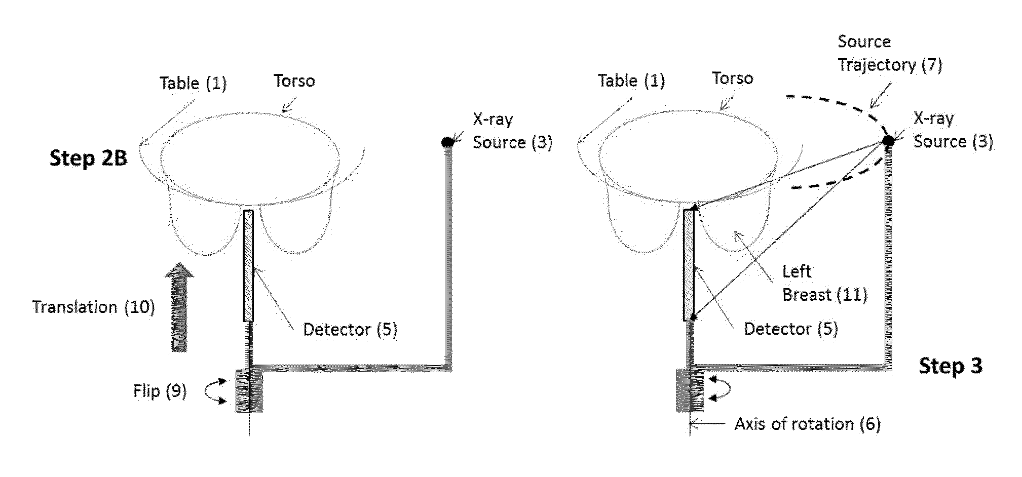
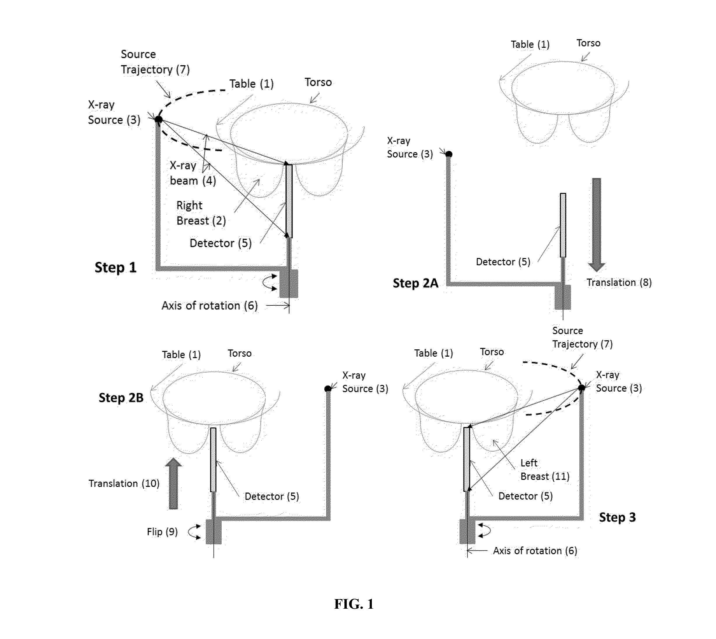
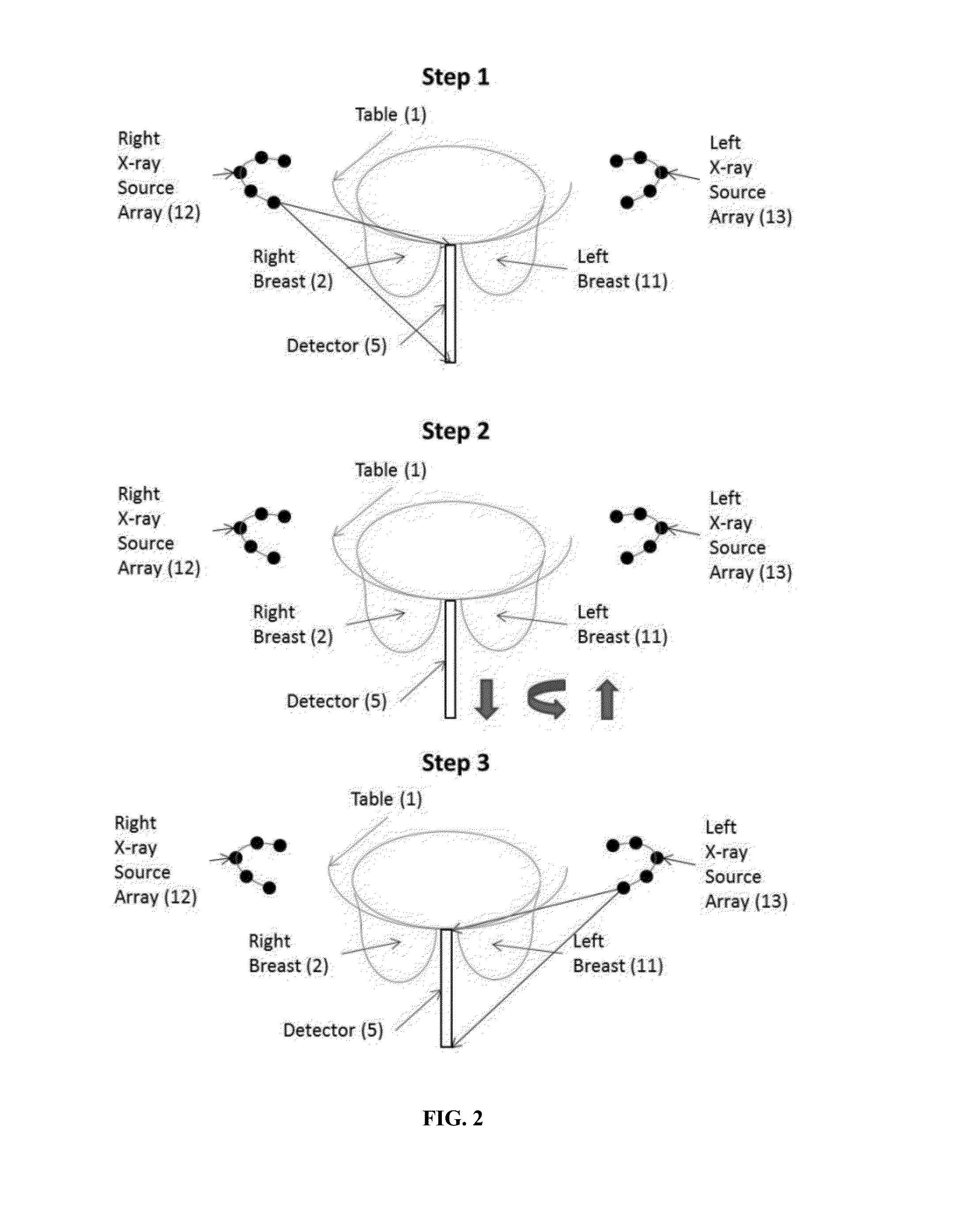
The invention is about x-ray-based breast imaging systems and methods that use a single detector and one or two x-ray sources to create high-quality images with improved spatial resolution. These systems can perform tomosynthesis or stereoscopic imaging without the need to reposition the patient during the procedure, making the process faster and more affordable.

Problems solved by technology

However, cost and access are major limitations for MRI tests.
Also, breast MRI scans have much lower spatial resolution than mammography.
Since mammography provides a 2-D image of the 3-D breast, superposition of structures leads to anatomic noise, which can mask lesions and / or mimic the presence of lesions that can lead to additional imaging and tissue biopsy.
However, mammography and digital breast tomosynthesis systems that are currently used provide only anatomic images of the breast.
A major limitation of existing systems for contrast enhanced digital mammography and contrast enhanced digital breast tomosynthesis require repositioning of the subject, as the system is designed to image one breast at a time.
This may result in the need for two injections that double the amount of injected contrast media, for bilateral exams.
This method provided a singular time-point on contrast enhancement and did not provide for studying contrast enhancement kinetics.
While it is possible to acquire multiple images at different time points after contrast administration, the temporal resolution of such an approach is limited by the frame-rate capabilities of the x-ray imaging detector.
Thus, the method takes 4.5 minutes to acquire the dataset that represents a single time point and hence suffers from substantial limitations on studying contrast enhancement kinetics.

Method used


Image

  • Apparatus and method for x-ray-based breast imaging
  • Apparatus and method for x-ray-based breast imaging
  • Apparatus and method for x-ray-based breast imaging

Examples


Embodiment Construction

[0034]The present invention provides unique x-ray breast imaging systems and related methods that are applicable to contrast enhanced digital mammography and contrast enhanced digital breast tomosynthesis. The invention enables fast, cost-effective and accurate x-ray imaging.

[0035]A unique aspect of the invention is the use of a single x-ray detector with the versatility of working in association with diverse x-ray source set-ups (e.g., one or two x-ray sources or arrays of x-ray sources) to generate fast and high-quality images, all without the need to reposition the patient during the imaging procedure. The invention is directly applicable to contrast enhanced digital mammography and contrast enhanced tomographic imaging such as digital breast tomosynthesis, which are considered as highly-promising candidates for clinical success. Moreover, the invention addresses limitations of existing systems and enables simultaneous acquisition of images for bilateral exams. Another exemplary ...


PUM


Abstract

The invention provides x-ray-based breast imaging systems and related methods that are, for example, applicable to contrast enhanced digital mammography and contrast enhanced digital breast tomosynthesis and allow fast, cost-effective and accurate x-ray imaging.

Description

PRIORITY CLAIMS AND RELATED PATENT APPLICATIONS[0001]This application claims the benefit of priority from U.S. Provisional Application Ser. No. 61 / 774,632, filed on Mar. 8, 2013, the entire content of which is incorporated herein by reference in its entirety.STATEMENT REGARDING FEDERALLY FUNDED RESEARCH OR DEVELOPMENT[0002]The United States Government has certain rights to the invention pursuant to Grant Nos. R21 CA134128 and R01 CA128906 from the National Institutes of Health to the University of Massachusetts.TECHNICAL FIELD OF THE INVENTION[0003]The invention generally relates to x-ray-based imaging systems and methods. More particularly, the invention relates to x-ray breast imaging systems and related methods that are, for example, applicable to contrast enhanced digital mammography and contrast enhanced digital breast tomosynthesis.BACKGROUND OF THE INVENTION[0004]Early detection and accurate diagnosis of breast cancer with screening and diagnostic mammography are critical asp...

Claims


Application Information

Patent Timeline
Patent Type & Authority Applications(United States)
IPC IPC(8): A61B6/00A61B6/02A61B6/04
CPCA61B6/502A61B6/40A61B6/4208A61B6/54A61B6/5211A61B6/485A61B6/481A61B6/4452A61B6/02A61B6/04A61B6/4007A61B6/461A61B6/025A61B6/032A61B6/4435A61B6/0435
Inventor VEDANTHAM, SRINIVASANKARELLAS, ANDREW
Owner UNIV OF MASSACHUSETTS
Learn More