Method and Apparatus for Repairing the Mid-Foot Region Via an Intramedullary Nail

a technology of intramedullary nail and midfoot, which is applied in the field of method and apparatus for repairing damaged, deteriorating, or fractured bones in the midfoot region, and can solve the problems of deteriorating the bones in the foot, limiting diabetic foot treatment to date, and collapse of the foo

Inactive Publication Date: 2016-06-09
HOWMEDICA OSTEONICS CORP
View PDF2 Cites 7 Cited by
  • Summary
  • Abstract
  • Description
  • Claims
  • Application Information

AI Technical Summary

Benefits of technology

[0045]Although prior art nails have found varying degrees of success in the treatment of long bone fractures and hind-foot arthrodesis, there remains a need in the field for an intramedullary nail specific for fixation of the medial column of the foot. This device will substantially reduce, if not eliminate many of the present complications encountered with Diabetic Charcot foot reconstruction. Such a device would provide fixation and stabilization of the Diabetic Charcot foot preventing future mid-foot collapse and ultimate limb loss. An intramedullary medial column nail and method of utilization would provide stabilization to the medial column of the foot, eliminating excessive operating time, anesthesia exposure, implant failure, long hospital stays, and reduce overall medical cost leading to early rehabilitation and ultimately limb salvage.
[0047]The present invention utilizes an intramedullary medial column nail fixation apparatus ideally being cylindrical and cannulated, and made from either titanium, a bioabsorbable material, human or animal bone, or equivalent. The nail ideally would have a number of proximal and distal fastener holes (transfixation or interlocking holes) for securing the IM medial column nail to the appropriate osseous cortical structures of the foot. An important feature of the present invention of the IM nail is its ability to function as an internal load-sharing device along the medial column of the foot. The nail acts as an internal splintage while the bones are mending, and is used to bring into alignment microfractures that may exist. The proximal portion of the nail may have a tapered tip to aid in insertion, and at least one fastener hole (ideally two) for the insertion of fasteners to fixate the neck of the talus to the implant or nail. The intermediate portion ideally has two slots to accommodate for any bone loss of the navicular and medial cuneiform, and to allow for the possible insertion of a fastener if the bones are healthy enough. The distal portion has at least one fastener hole (ideally three) for the insertion of fasteners to fixate the nail to the first metatarsal.
[0049]In addition, another embodiment of the present invention calls for the use of a compression system that incorporates a depth gauge device which may be used to determine the amount of compression achieved. This device would eliminate the need for guessing or “feeling” the correct amount of compression required. A depth gauge would exist either on the implant or jig apparatus with a ruler type measuring means for gauging the amount of travel in the nail back up the intramedullary canal towards the phalanges. Compression could then be determined to a specified length of travel of the nail back up the canal during compression.

Problems solved by technology

Diabetic neuropathy ultimately results in deterioration of the bones in the foot.
Such deterioration of those bones in the mid-foot region leads to the collapse of the foot, fracture of those bones, or both.
Diabetic foot treatment to date has been limited to amputation, special casts or shoes which alleviate the deformity, and surgical intervention to strengthen the foot.
Surgical intervention has been restricted to the use of screws and plates since most bones in the foot are quite small.

Method used


Image

  • Method and Apparatus for Repairing the Mid-Foot Region Via an Intramedullary Nail
  • Method and Apparatus for Repairing the Mid-Foot Region Via an Intramedullary Nail
  • Method and Apparatus for Repairing the Mid-Foot Region Via an Intramedullary Nail

Examples


Embodiment Construction

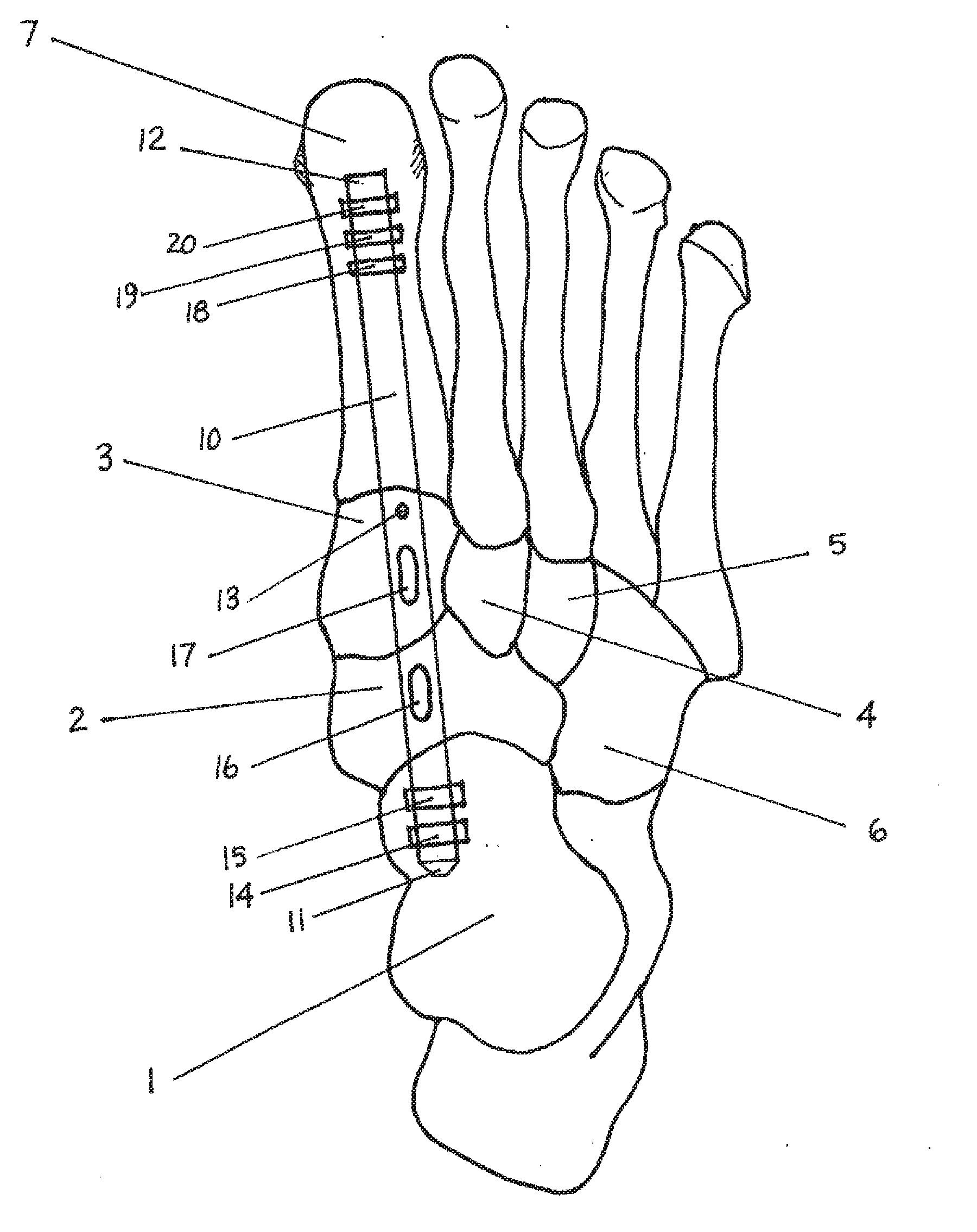
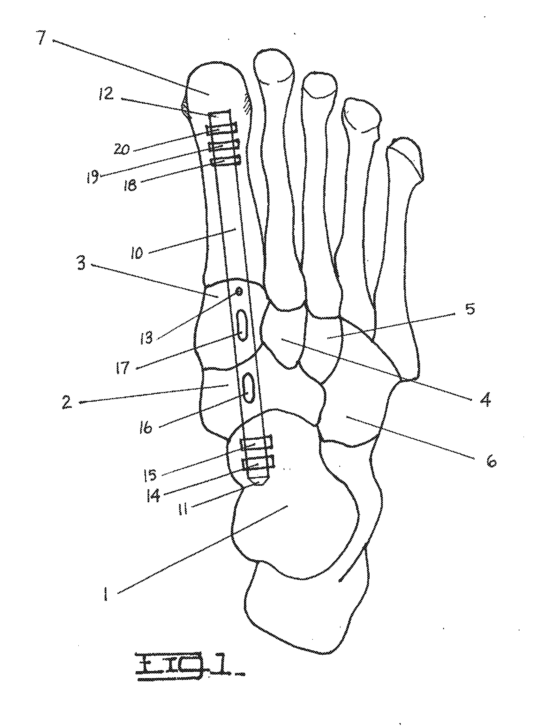
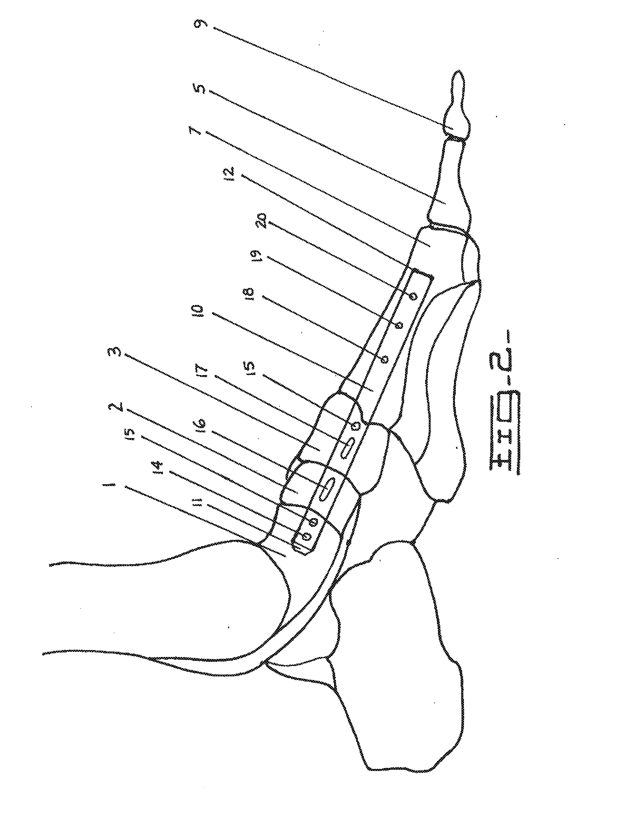
[0053]The following description will explain the embodiments of the invention, beginning with a device used for treating deteriorating or damaged bones in the medial column of a human foot, and concluding with the method for treating deteriorating or damaged bones in the medial column of a human foot. Both the first and second figures are skeletal outlines of a human foot, depicting the device therein and the resulting orientation of the bones by using the method specified below.

[0054]FIG. 1 is a top down view of a right human foot, depicting the bones therein and an implant or intramedullary nail 10 running through the mid-foot region. The implant or intramedullary nail 10 runs through the medullary canal of first metatarsal 7, medial cuneiform 3, navicular 2, and talus bone 1. The talus bone 1 makes up the lower part 20 of the ankle joint where the proximal end 11 of the implant or intramedullary nail 10 is attached with at least one fastener (or locking screw, with two depicted i...


PUM


Abstract

A device, method, and system for treatment or fixation of a fractured, damaged, or deteriorating bone or bones in a mid-foot region. The device comprising an implant with both proximal and distal fastener holes, along with fastener slots in a central elongated body, for securing the implant to the appropriate osseous cortical structures of the foot. The method for treatment or fixation of fractured, damaged, or deteriorating bones in the medial column of the foot with use of a device such as an intramedullary nail that attaches to either the talus or first metatarsal bones to secure the medial cuneiform and navicular bones in place.

Description

PRIORITY[0001]Applicant hereby claim priority under all rights to which they are entitled per 35 USC 120 based upon the U.S. Application No. 60 / 402,380 for this Patent Cooperation Treaty (PCT) patent application (USPTO receiving office) filed at the United States Patent and Trademark Office on Aug. 10, 2002. This application also takes priority from PCT / US2003 / 024185 entitled, “Method and Apparatus for Repairing the Mid-Foot Region Via an Intramedullary Nail”, filed on Aug. 1, 2003 and its corresponding US National Stage application under 35 USC 371, Ser. No. 10 / 525,544 of the same title and filed Jan. 28, 2005. Priority is also claimed to the corresponding continuation application Ser. No. 13 / 593,987 of the same title filed on Aug. 24, 2012. The entire contents of the above referenced applications are also herewith incorporated by reference, in their entirety, in the present application.BACKGROUND[0002]1. Field of the Invention[0003]The field of the present invention relates to a m...

Claims


Application Information

Patent Timeline
Patent Type & Authority Applications(United States)
IPC IPC(8): A61B17/72
CPCA61B17/7216A61B17/7291A61B17/72A61B17/7225A61B17/7233A61B17/8897A61B17/7241A61B2017/681A61B17/846
Inventor SIMON, WILLIAM H.
Owner HOWMEDICA OSTEONICS CORP
Why Eureka
Try Eureka