Simulated tissue structures and methods

a tissue structure and tissue technology, applied in the field of surgical training tools, can solve the problems of difficult to achieve, reduce the realism arising from compensation, damage to the outer geometry of geometry, etc., and achieve the effect of reducing realism and being easy to remove from the mandrel

Active Publication Date: 2016-10-06
APPL MEDICAL RESOURCES CORP
View PDF0 Cites 44 Cited by
  • Summary
  • Abstract
  • Description
  • Claims
  • Application Information

AI Technical Summary

Problems solved by technology

Also, the internal material has a different and more rigid characteristic relative to the external material and the anatomical geometry formed by the outer material would normally make insertion of the internal material into the external geometry damaging to the outer geometry, difficult to accomplish and would reduce the realism arising from compensations, such as repairing and gluing, necessary due to damaging insertion.

Method used


Image

  • Simulated tissue structures and methods
  • Simulated tissue structures and methods
  • Simulated tissue structures and methods

Examples


Embodiment Construction

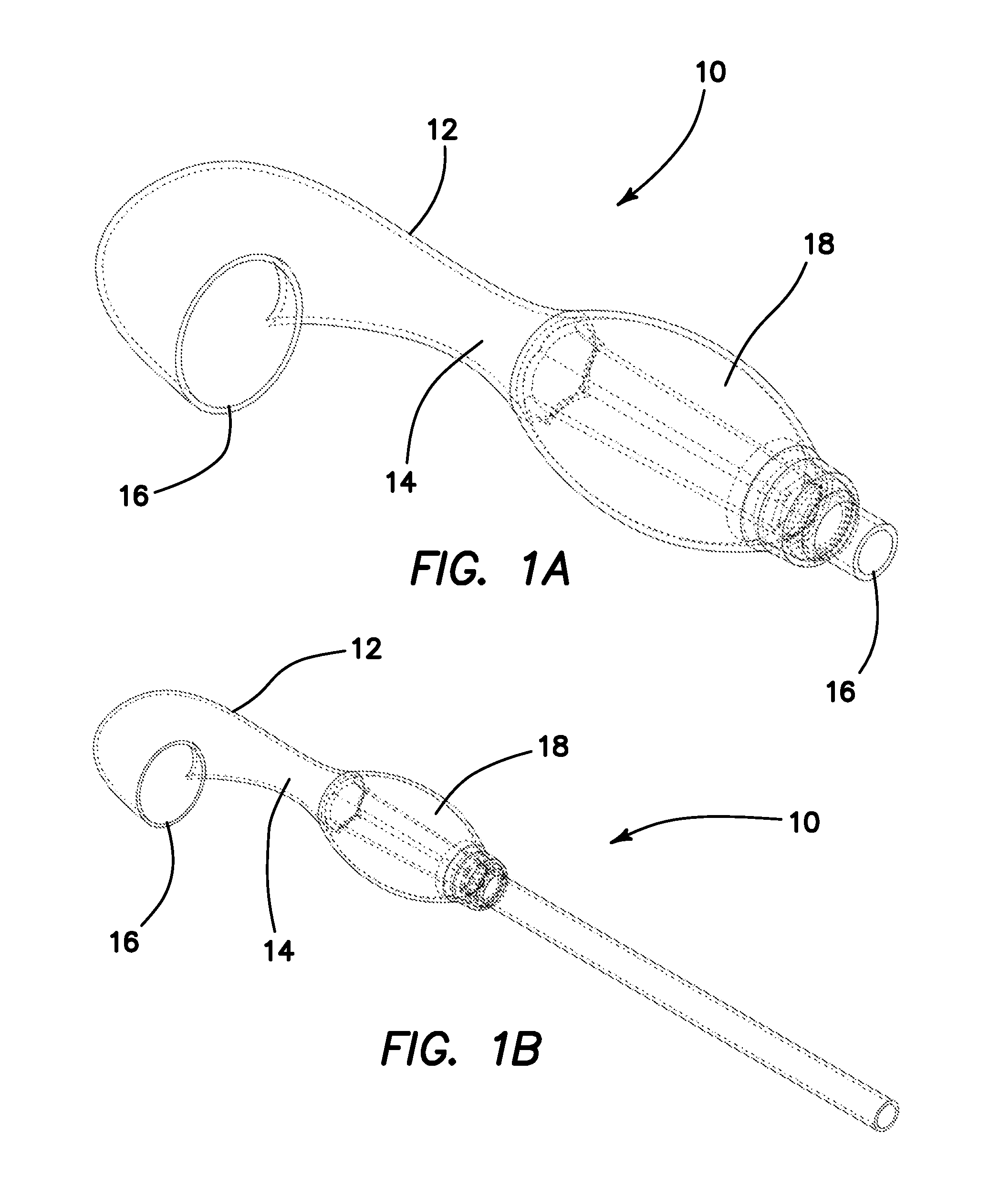
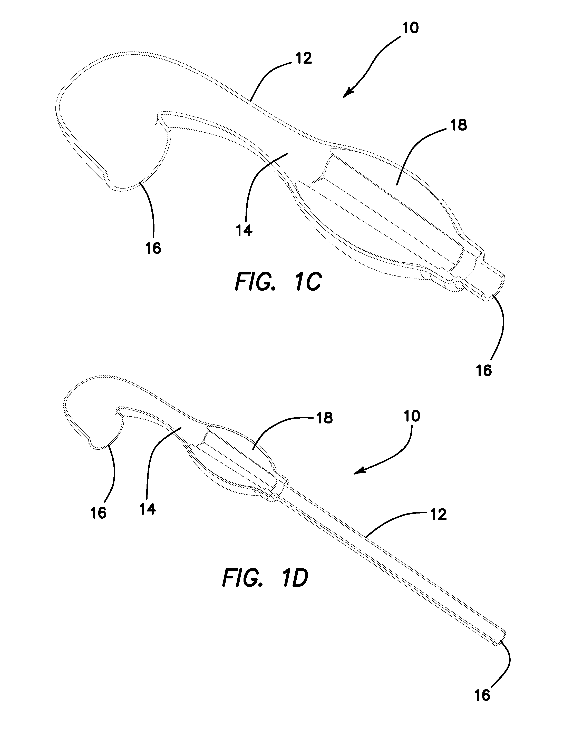
[0068]Turning now to FIGS. 1A-1D, there is shown a simulated tissue structure 10 according to the present invention. The simulated tissue structure 10 includes a silicone outer portion 12 having an outer surface and an inner surface. The inner surface defines an interior cavity 14. The interior cavity 14 is interconnected with at least one opening 16. The cavity 14 of the simulated tissue structure 10 of FIGS. 1A-1D includes two openings 16 and the cavity 14 is lumen-like and generally elongated. In particular, the outer portion 12 is configured to have a size and shape of a tissue structure, organ, or at least a part of an anatomy. For example, as shown in FIGS. 1A-1D, the outer portion 12 is configured in shape and size to represent a fallopian tube of the female human anatomy. FIGS. 1B and 1C illustrate a proximal elongation that is longer so as to integrally form a fallopian tube than shown in FIGS. 1A and 1C so as to be optionally connectable to a separately formed fallopian tu...


PUM

PropertyMeasurementUnit
diameteraaaaaaaaaa
tissue structureaaaaaaaaaa
lengthaaaaaaaaaa

Abstract

Simulated tissue structures and methods of manufacturing are provided. The simulated tissue structures are particularly useful for placement inside abdominal simulators for practicing laparoscopic surgical techniques. One simulated tissue structure includes a combination of two materials that are attached together wherein one of the materials forms a hollow anatomical structure configured to contain the other material. The two materials are attached in an anatomically advantageous manner such that the inner surface of the outer material closely conforms to the outer surface of the inner material. Another simulated tissue structure includes a plurality of layers wherein at least one layer is applied by printing the layer with at least one stencil to impart one or more functional characteristic to the simulated tissue structure.

Description

CROSS-REFERENCE TO RELATED APPLICATIONS[0001]This application is a continuation of International Application No. PCT / US2016 / 018697 entitled “Simulated tissue structures and methods” filed on Feb. 19, 2016 which claims priority to and benefit of U.S. Provisional Patent Application No. 62 / 118,179 entitled “Method of making simulated tissue using stencils” filed on Feb. 19, 2015 and U.S. Provisional Patent Application No. 62 / 119,542 entitled “Foam-on-mandrel method of making simulated anatomy” filed on Feb. 23, 2015 all of which are incorporated herein by reference in their entireties.FIELD OF THE INVENTION[0002]This application relates to surgical training tools, and in particular, to simulated tissue structures and organ models for teaching and practicing surgical procedures and methods for making them.BACKGROUND OF THE INVENTION[0003]A highly-skilled operation technique is required of surgeons, in general, and, in particular, for performing laparoscopic surgical procedures. In lapar...

Claims


Application Information

Patent Timeline
Patent Type & Authority Applications(United States)
IPC IPC(8): G09B23/28G09B23/30
CPCG09B23/30G09B23/281B29L2031/7532B29L2031/753B29C41/20G09B23/28G09B23/34
Inventor HOFSTETTER, GREGORY K.
Owner APPL MEDICAL RESOURCES CORP
Why Eureka
Try Eureka