Automatic segmentation of breast tissue in a thermographic image

a technology of breast tissue and thermographic image, which is applied in the direction of image enhancement, medical/anatomical pattern recognition, instruments, etc., can solve the problems of difficult management of one or more regions of interest in a thermographic image, and the challenge of extracting one or more regions of interes

Active Publication Date: 2017-08-31
NIRAMAI HEALTH ANALYTIX PVT LTD
View PDF8 Cites 2 Cited by
  • Summary
  • Abstract
  • Description
  • Claims
  • Application Information

AI Technical Summary

Benefits of technology

[0004]What is disclosed is a system and method for automatically segmenting breast tissue from surrounding tissue in a thermal image of a patient for breast cancer screening. One embodiment of the present method involves the following. First, a thermographic image of at least one breast of a person undergoing breast cancer screening is received. Pixels in the thermographic image with a higher temperature value are displayed in a first color and pixels with a lower temperature value are displayed in a second color. Pixels with temperature values between the lower and higher temperature values are displayed in gradations of color between the first and second colors. The thermographic image is then analyzed to identify a set of N≧5 points around the breast based on a contour of an outline of the body and the isotherms of the axilla and infra-mammary fold. Thereafter, the points are connected, in a piecewise linear fashion, to form a N-sided polygon which segments the breast from surrounding tissue. Each of the points is a vertex of the polygon and comprises a draggable object which enables a user to selectively manipulate a shape of the polygon. The teachings hereof find their uses in a breast cancer screening software tool.
[0005]Features and advantages of the above-described method will become readily apparent from the following detailed description and accompanying drawings.

Problems solved by technology

Extraction of one or more regions of interest in a thermo graphic image of is a challenging task due to the amorphous nature of the tissue undergoing examination.
Although experts can segment the breast tissue into regions of interest by visual inspection after years of experience, it becomes a difficult task to manage effectively when the number of patients being screened is larger than a few.

Method used


Image

  • Automatic segmentation of breast tissue in a thermographic image
  • Automatic segmentation of breast tissue in a thermographic image
  • Automatic segmentation of breast tissue in a thermographic image

Examples


Embodiment Construction

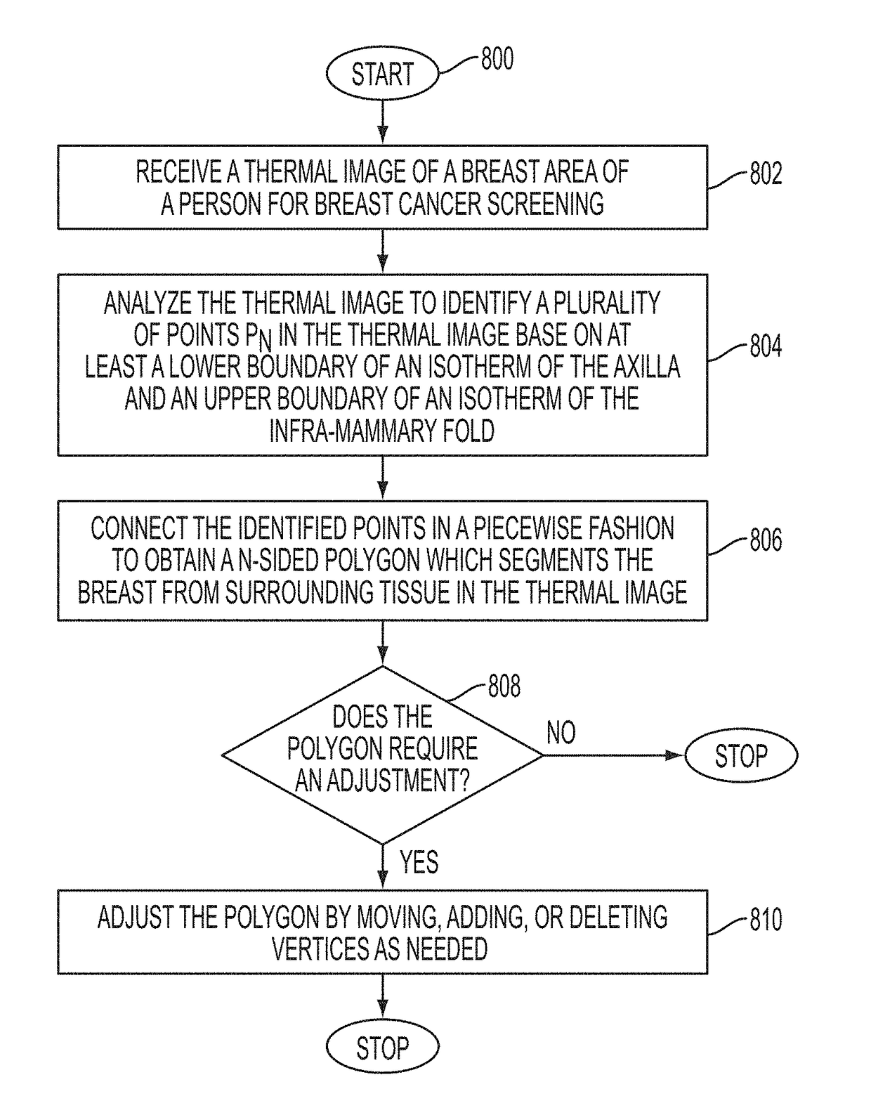
[0017]What is disclosed is a system and method for automatically segmenting breast tissue from surrounding tissue in a thermal image of a patient for breast cancer screening. The teachings hereof find their intended uses in a software interface tool performing automated or semi-automated breast cancer screening.

[0018]A “person” refers to either a male or a female. Gender pronouns are not to be viewed as limiting the scope of the appended claims strictly to females. Moreover, although the term “person” or “patient” is used interchangeably throughout this disclosure, it should be appreciated that the person undergoing breast cancer screening may be something other than a human such as, for example, a primate. Therefore, the use of such terms is not to be viewed as limiting the scope of the appended claims to humans.

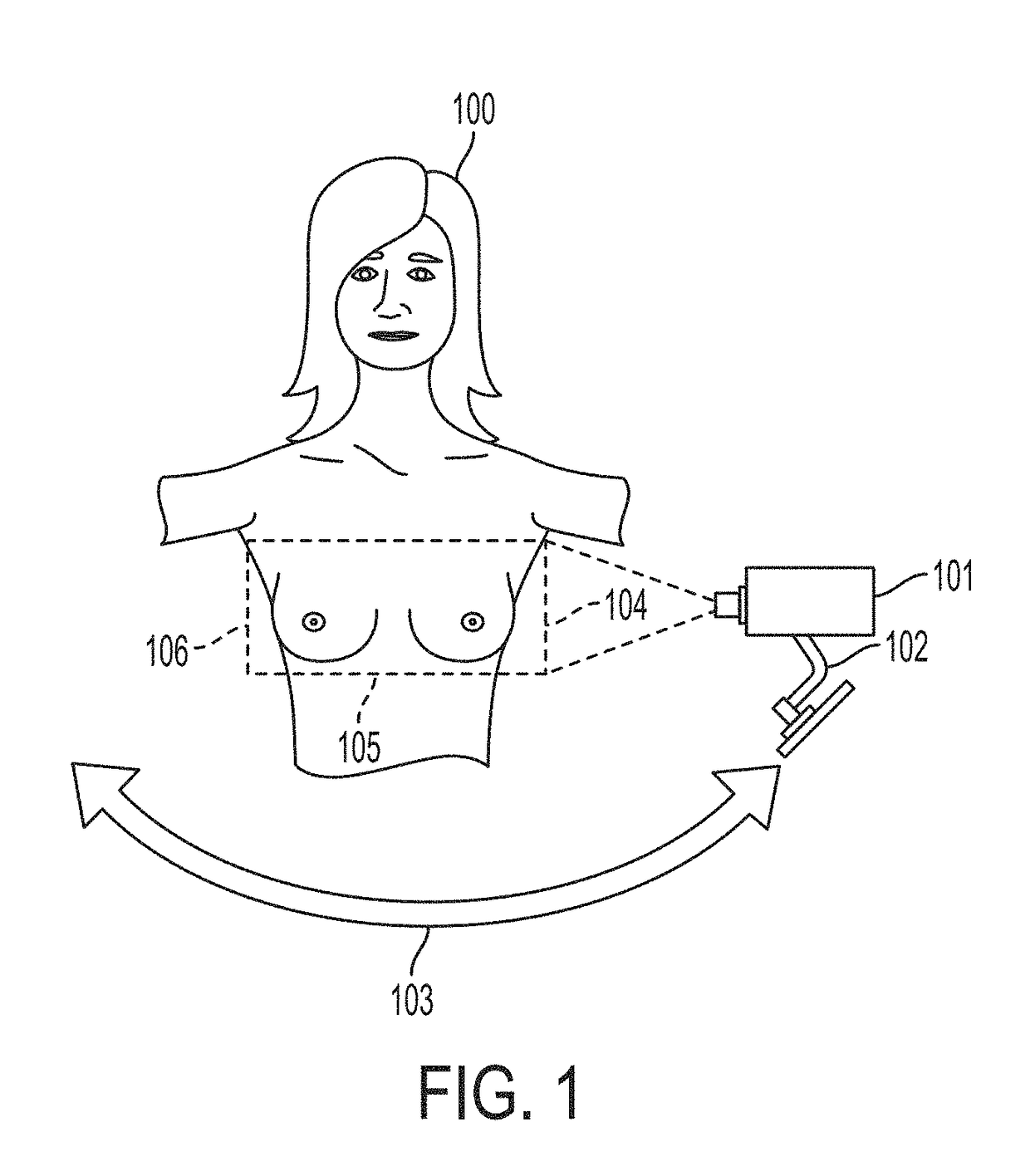
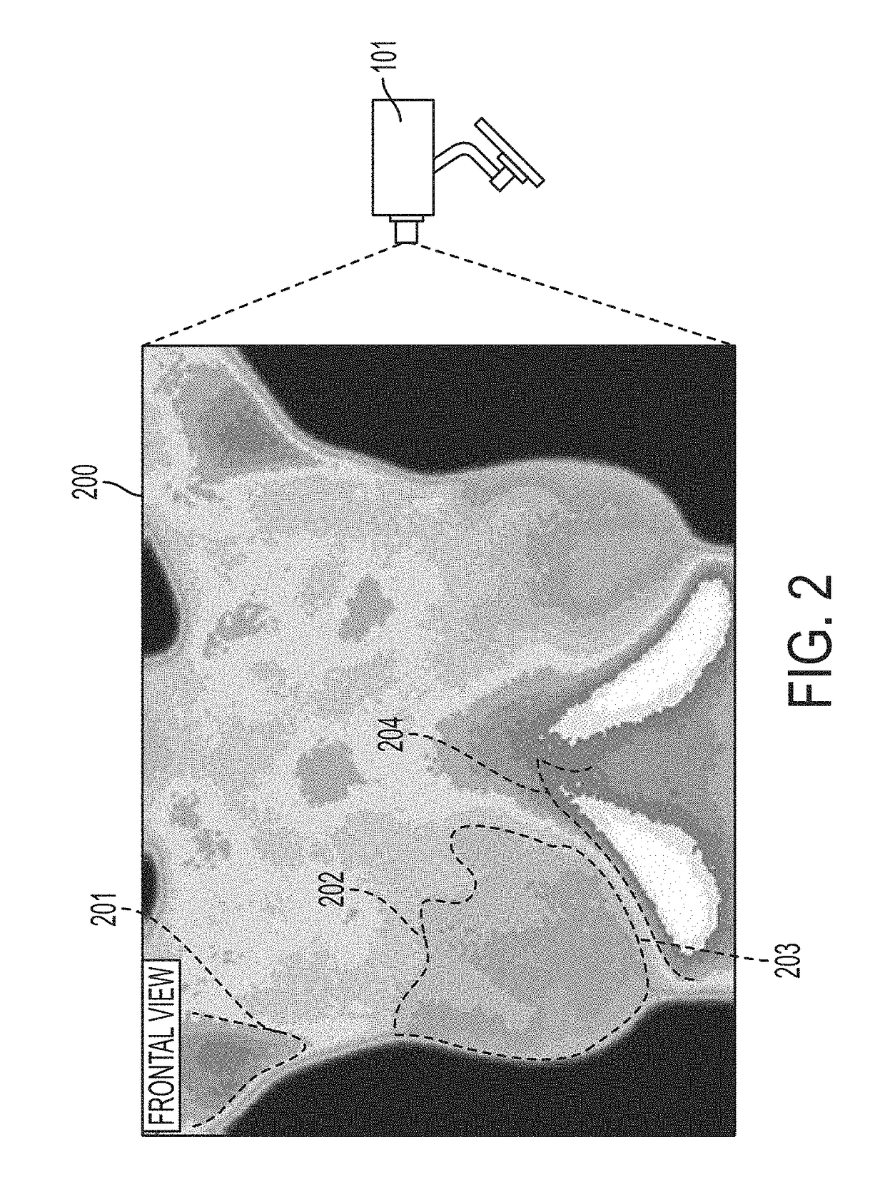
[0019]A “breast area” refers to tissue of the breast and may further include surrounding tissue as is deemed appropriate for breast cancer screening. Thermal images are cap...


PUM


Abstract

What is disclosed is a system and method for automatically segmenting a breast from surrounding tissue in a thermal image. A thermal image of at least one breast of the patient is received. The thermal image is then analyzed to identify a set of N points around the breast, a contour of an outline of the body, and isotherms of the axilla and infra-mammary fold. Thereafter, the points are connected together to form a N-sided irregular polygon which segments the breast from surrounding tissue. Each of the points is a vertex of the polygon and comprises a draggable object which enables a user to selectively manipulate a shape of the polygon. A user can add/delete vertices from the polygon as desired. The area of the image encompassed by the polygon is communicated to a breast cancer screening algorithm performing automated or semi-automated screening.

Description

TECHNICAL FIELD[0001]The present invention is directed to systems and methods for automatically segmenting breast tissue from surrounding tissue in a thermal image of a patient for breast cancer screening.BACKGROUND[0002]Automated thermographic screening can be very useful and effective for conducting breast cancer screening where knowledge and expertise of the specific Region of Interest (ROI) of the breast is required to separate a specific region of interest from surrounding tissues which are of no interest so that the identified tissue can be further analyzed and subsequently classified as being cancerous or non-cancerous. Extraction of one or more regions of interest in a thermo graphic image of is a challenging task due to the amorphous nature of the tissue undergoing examination. Although experts can segment the breast tissue into regions of interest by visual inspection after years of experience, it becomes a difficult task to manage effectively when the number of patients b...

Claims


Application Information

Patent Timeline
Patent Type & Authority Applications(United States)
IPC IPC(8): G06T7/00G06T7/60A61B10/00G06K9/46A61B5/00A61B5/01G06T7/40G06K9/52G06V10/44G06V10/56G06V20/00
CPCG06T7/0012G06T7/408G06T7/60G06T7/0085G06K9/52G06T2207/10048G06K9/4604A61B5/0091A61B5/015A61B10/0041G06T2207/30068G06K9/4652A61B5/4312A61B2576/02G06T7/90G16H30/40G06V20/64G06V20/00G06V10/44G06V10/56G06V2201/03G06T7/13
Inventor PARTHASARATHY, ARUN KOUSHIKVENKATARAMANI, KRITHIKAKAKILETI, SIVA TEJA
Owner NIRAMAI HEALTH ANALYTIX PVT LTD
Why Eureka
Try Eureka