Three-dimensional MRI image-based breast tumor automatic segmentation method

A breast tumor, automatic segmentation technology, applied in the field of medical image processing, to save time, reduce workload, and achieve high segmentation accuracy

Active Publication Date: 2017-12-12
THE SECOND PEOPLES HOSPITAL OF SHENZHEN
View PDF8 Cites 45 Cited by
  • Summary
  • Abstract
  • Description
  • Claims
  • Application Information

AI Technical Summary

Problems solved by technology

[0004] The purpose of the present invention is to provide a method for automatic segmentation of breast tumors based on three-dimensional MRI images, aiming to solve the problem that in general, t

Method used


Image

  • Three-dimensional MRI image-based breast tumor automatic segmentation method
  • Three-dimensional MRI image-based breast tumor automatic segmentation method
  • Three-dimensional MRI image-based breast tumor automatic segmentation method

Examples


Embodiment Construction

[0012] In order to make the technical problems, technical solutions, and beneficial effects to be solved by the present invention clearer, the following further describes the present invention in detail with reference to embodiments. It should be understood that the specific embodiments described herein are only used to explain the present invention, but not to limit the present invention.

[0013] The embodiment of the present invention provides a method for automatically segmenting breast tumors based on three-dimensional MRI images, which includes the following steps:

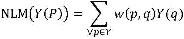
[0014] S01. Image preprocessing: provide an initial MRI image, and preprocess the initial MRI image using a non-local average filter;

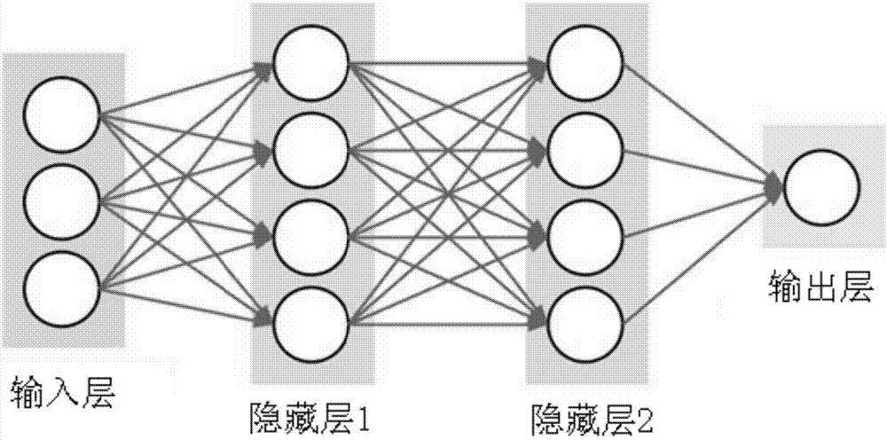
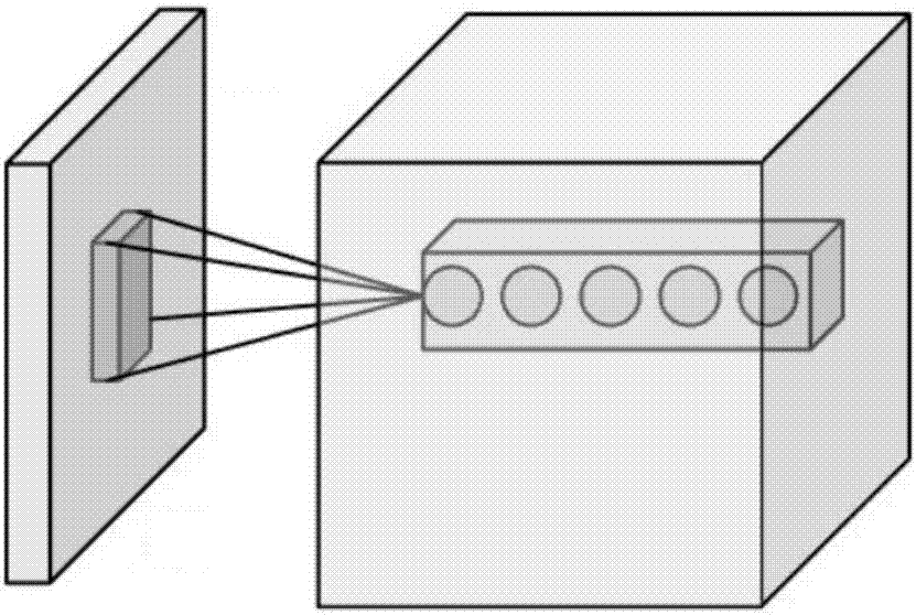
[0015] S02. Breast tumor location: build a multi-layer processing model on the training set, use convolutional neural network to abstract the features of the training object, automatically extract segmentation features, and output the probability distribution map of tumor location;...


PUM


Abstract

The invention provides a three-dimensional MRI image-based breast tumor automatic segmentation method. The method comprises the following steps of performing image preprocessing: providing an initial MRI image and performing preprocessing on the initial MRI image by adopting a non local mean filter; performing breast tumor locating: building a multilayer processing model for a training set, performing hierarchical abstraction on characteristics of a training object by adopting a convolutional neural network, automatically extracting segmentation characteristics, and outputting a probability distribution graph of a tumor position; performing breast tumor boundary segmentation: providing a breast three-dimensional MRI image, determining a seed point based on the probability distribution graph of the tumor position, and finishing segmentation initialization to obtain an initial region C0 of a tumor; and performing accurate segmentation on the tumor by using a region growth algorithm.

Description

Technical field [0001] The invention belongs to the field of medical image processing, and in particular relates to a method for automatically segmenting breast tumors based on three-dimensional MRI images. Background technique [0002] Breast cancer is the most common malignant tumor among women, and its fatality rate ranks first among female malignant tumor death rates worldwide. Currently, early diagnosis and timely treatment are the most effective measures to deal with breast cancer. Medical imaging methods, such as X-ray, magnetic resonance imaging (MRI), and ultrasound, are currently the most important methods for detecting and diagnosing breast cancer. Among them, MRI can better distinguish various organizations and provide doctors with sufficient reference information. However, in general, doctors need to manually segment breast tumors, which takes a lot of time and energy, and the diagnosis accuracy is affected by the doctor's professional ability and experience subjec...

Claims


Application Information

Patent Timeline
IPC IPC(8): G06T7/12
CPCG06T2207/10088G06T2207/20081G06T2207/20084G06T2207/30068G06T2207/30096G06T7/12
Inventor 林帆雷益
Owner THE SECOND PEOPLES HOSPITAL OF SHENZHEN
Why Eureka
Try Eureka